Microscopes used for microphotographs presented on this site
By clicking on each of the images below, representing the different microscopes I’ve used, you’ll land on a page on which you can find some informations about the microscope, its main characteristics and annotated photomicrographs.
Each microscope brings different informations : on samples surfaces for scanning electron microscope and atomic force microscope; on internal structure of organs for light microscope and electron transmission microscope.
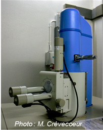
Electron Transmission Microscope FEI TECNAI TM G2 Sphera
Sciences II - Bioimaging Center
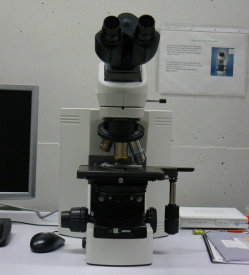
Optical Microscope Nikon Eclipse 80i
Botany and Plant Biology
Sciences III
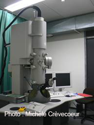
Scanning Electron Microscope JEOL JSM 6510LV
Sciences II - Bioimaging Center
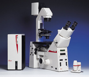
Optical Microscope Leica DMI – RE 2
Botany and Plant Biology
Sciences III
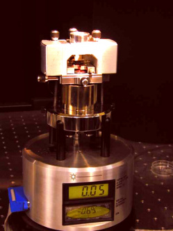
Atomic Force Microscope Nanoscope III
Prof. E. Lesniewska Laboratory
University of Bourgogne - Dijon - France
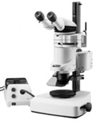
Steoroscopic Microscope Leica MZ 16
Botany and Plant Biology
Sciences III
Microscopy University of Geneva. Bioimaging Center Sciences Faculty (Sciences II) – University of Geneva
Bio-Imaging Core Facility Microscopie photonique pour l’observation de cellules vivantes ou fixées. CMU – University of Geneva