The Atomic Force Microscope designated by the abbreviation « AFM » belongs to the family of near field microscopes or local probes microscopes. It is used to obtain high resolution images, on the nanometer scale, of the surface of biological samples. It allows to observe and image in air samples but also in liquid, physiological conditions. In addition, samples viewed by AFM do not need to be conductors and do not require any preparation that would change or damage the sample. The resolution of AFM is high: lateral 0,5 -1 nm lateral; vertical 0,01 nm lateral for dry samples and 0,1 – 0,2 nm for samples imaged in physiological conditions. AFM can not only give image in the 3 D topography, but it also provides qualitative and quantitative information and different physical properties (surface texture and roughness; size).
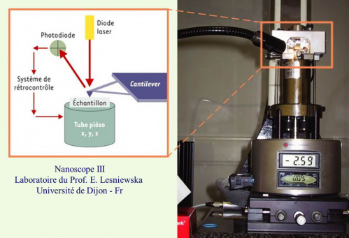
A sharp tip attached to a cantilever is scanned over the sample surface and moves in response to tip – surface interactions at the atom level, the movement being measured with a laser beam photodiode. Different forces are involved in the imaging process.
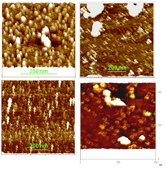
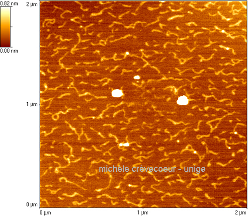
Below a few AFM pictures of (1) purified membranes from spinach leaves observed in buffer and (2) polygalacturonic acid molecules observed in air after drying (protocol for preparation in French).
See the pdf Document in French » AFM en biologie » – cours de Microscopie électronique – M. CREVECOEUR UniGe
Membranes from spinach leaf cells imaged by AFM See the publication « Crèvecoeur M et al. (2000) – Protoplasma 212: 46-55 [pdf] »
Spinach plasma membranes have been imaged with a Nanoscope III microscope - laboratory of Prof. Eric Lesniewska - University of Burgoundi - Dijon - France
Polygalacturonic acid molécules observed dry on mica