Anatomy of vegetative organs and of flower buds of angiosperms,
both monocotyledons and dicotyledons.
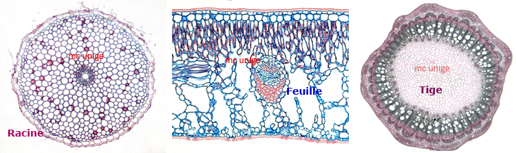
This part of the site, devoted to plant anatomy, presents microphotographs of sections through different organs from monocotyledons and dicotyledons. (see below).
Some notions relative to morphology of vegetative organs will be found on another page.
Superior plants, unable to move, have had to develop the ability to live in a wide variety of environments, sometimes extreme. This is reflected among other things by the astonishing plasticity of their vegetative organs’ morphylogy. The study of the tissues constituting these organs has proved to be an essential tool for the study of this great morphological plasticity and for the comparison of different phenotypes, wild and mutant (see Research).
Most of micrographs concern fresh sections, made either with a Leica VTs Vibratome or with a razor blade and paraffin sections made with a rotary microtome, either Anglia or MicroTec cut 45 (see Instruments).
The most current staining’s (see Protocols) of sections used were :
Astra Blue – Basic Fuchsin; Carmin – green iodide for vibratome sections.
Toluidin blue; Alcian Blue – Safranin; Astra Blue – Basic Fuchsin for paraffin sections.
Some micrographs relate to Epon sections stained either with methylene blue – basic fuchsin or with toluidin blue and this will be indicated on the relevant page.
The micrographs were taken for the majority with a Nikon Eclipse 80i microscope equipped with a Nikon DigitaL Sight DDS color camera.
As far as possible, the scale, the type of section and the staining are indicated for each micrograph. If it is not it is because I have not been able to find this information in my archives…it should be done later I hope !
Finally, some micrographs illustrate sections through organs of plants with medicinal properties. The microscope slides come from the collection of Georges Roux – pharmacist in Alger. They were entrusted to me by his grand-daughter Céline Roux. For these, unfortunately information on the type of section is not available and the staining is not always indicated. It should be noted the year of their realization is indicated for some of them and it is about 1940. The state of their conservation is excellent. Micrographs were taken with a Leica DM 750 Microscope equipped with a Leica MC179HD camera.
The organs for which sections has been made is given here below.