Plant tissues classified according to their function
This part of the web site presents micrographs illustrating the different plant tissues. For each of the tissue indications are given on the method used for their characterization.
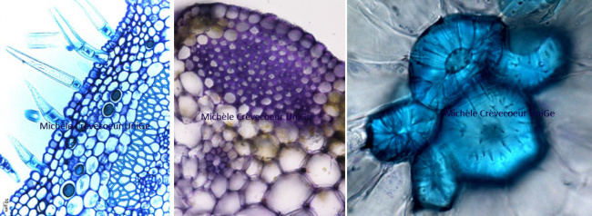
The classification of plant tissues can be made based on different criteria in particular the development, the function and the types of cells. On the basis of their function, we can distinguish 6 types of tissues listed here below.
1/ Dermal tissues: epidermis, periderm, rhizodermis (see root anatomy) and their specialized cells (stomata, trichomes)
2/ Ground tissue: parenchyma
3/ Supporting tissue: collenchyma and sclerenchyma (modified ground tissues)
4/ Conducting : primary xylem and phloem (see cross sections through roots and stems & longitudinal sections through stems) ; secondary xylem and phloem (wood and liber).
5/ Secretory & particular cells with crystals
6/ Meristematic: primary & secondary meristems; pericycle.
The study of plant tissues at light and electron microscopy includes a great variety of methods and techniques. The methodology used will vary depending on what we want to study. There are different methods for embedding and sectioning samples (paraffin, Epoxy resins, acrylic resins) and many staining protocols of sections. Information on the tissues can be sometimes obtained with very simple protocol and without equipment: fresh sections made using a razor blade can be stained directly and observed under a light microscope. Whatever the method used, simple or complex, it is possible to determine whether a section has been made through a root or a stem and if it corresponds to an organ from a monocotyledon or a dicotyledon. It is also possible to compare tissues from mutant and wild type plants and to study tissues from genetically modified plant.