Xylem and phloem in young stems Aristolochia rotunda, Zea mays et Asparagus.
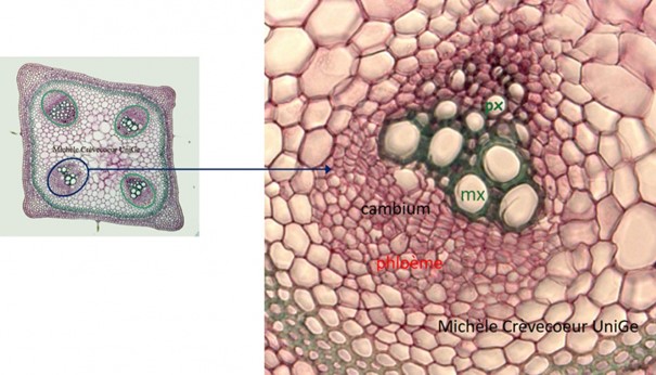
In young stems, both dicotyledons and monocotyledons, xylem and phloem are collateral with xylem towards center of the organ and phloem towards outside. This is clearly seen on cross sections as illustrated below. 1/ Example in a young stem of Aristoloche (dicotyledon). On the left detail of a vascular bundle on which this distribution is clearly seen. At the xylem level we distinguish the protoxylem (px) that consists of vessels of small diameter at the center and metaxylem (mx) composed of larger vessels. Their distribution has the appearance of an isosceles triangle with the protoxylem oriented toward the pith. See also the page : Aristolochia r. stem
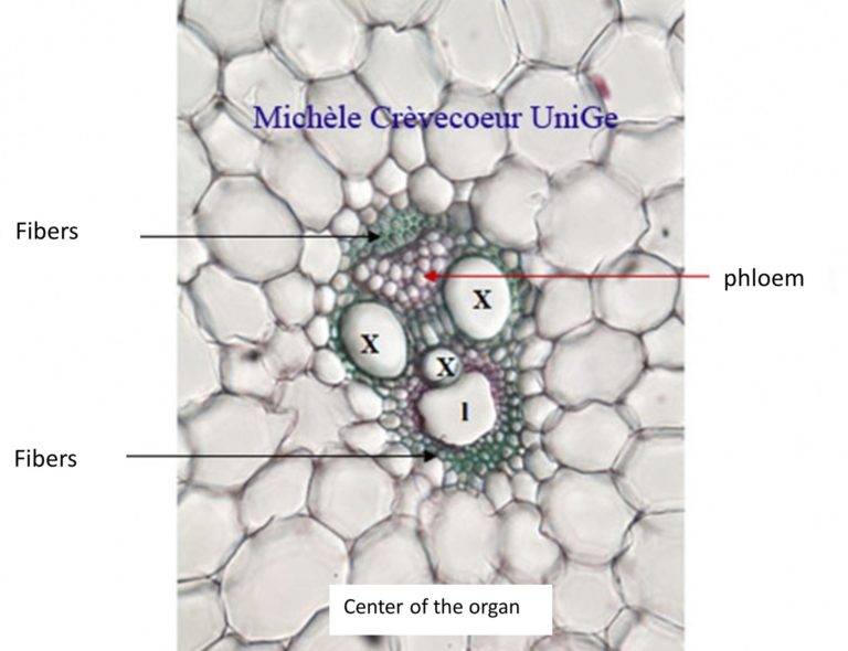
2/ Example in a young of Zea mays a monocotyledon.The superposition of xylem and phloem is less evident due to the V distribution of xylem and the phloem between arms formed by xylem (X). A few fibers (green) are observed at both extremities of the bundle. As result of elongation of internode, the most centered xylem is distended, and its primary wall disappears. This results in formation of a lacuna (l). See also the page : » Stem of Zea mays« .
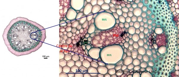
The distribution of primary xylem and phloem is also illustrated on a cross section in a stem of Asparagus, another monocotyledon. As in Zea mays stem a V distribution of xylem is observed, two large xylem vessels , the metaxylem (mx) vessels located towards the periphery of the organ outside and the protoxylem (px) vessels are located towards center. See also the page : Asparagus stem