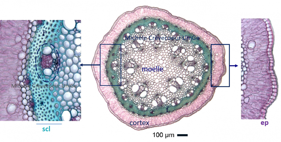
Microphotographs of a cross section through an Asparagus stem.
In this section tissues with cellulosic cell walls are stained pink and tissues with lignified cell walls are stained green.
Figure below : In the middle general aspect of the cross section. From outside towards inside we successively find: the monolayer of epidermis; the cortical parenchyma (cortex); a green ring of cells with lignified walls corresponding to the sclerenchyma; two rings of vascular bundles distributed in the pith (parenchyma).
On the left detailed portion of a vascular bundle with a V distribution of xylem and the phloem composed of small cells with cellulosic wall, between arms of this V.
On the right detail of the protective tissue, the epidermis with a thin cuticle and of the cortex.
Characteristics of this section:
Vascular bundles disposed on different rings (see the central picture with 2 apparent rings); colateral vascular bundles with V distribution of xylem; xylem and phloem superposed, two characteristics indicating that the section corresponds to a monocotyledon stem. For details see the page vascular tissues of stems.
The sclerenchyma (scl) forms a continuous ring of a few layers of fibers between cortical and pith parenchyma given support to the young stem.