Microphotographs of cross sections through a Zea mays stem

Two kinds of sections are shown below.
1/ The first ones are fresh cross sections made either with a razor blade or a vibratome in fresh stem portion. They have been stained with methyl green.
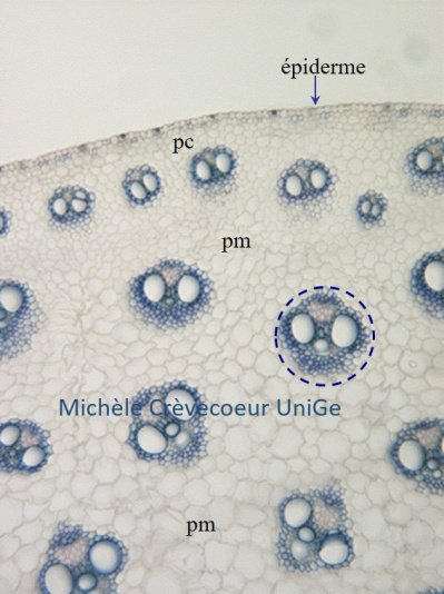
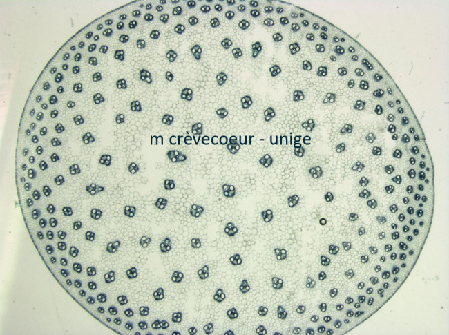
General aspect of the section shows scattered vascular bundles distributed on different rings inside the ground parenchyma (medular parenchyma) which occupy most part of the section. The cortex, that consists in cortical parenchyma, is reduced to a few rows of cells located below the epidermis and the first row of vascular bundles.

Miniphoto tige Zea mays

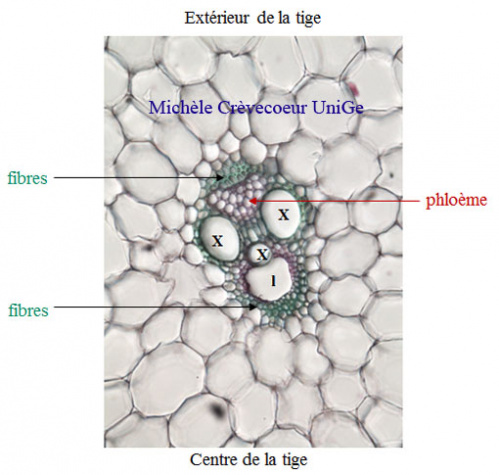
Below: part of a cross section through a young Zea mays stem with a detail of a vascular bundle on the right.1/

pm: medullar parenchyma; pc: cortical parenchyma 
The epidermis consists in a monolayer of cells with a very thin cuticle

Vascular bundle

The vascular bundles are colateral wtith superposed primary xylem (x) and primary phloem. The xylem shows a V distribution with phloem between arms of this V. Each vascular bundle is surrounded by fibers on both extremities. l: lacuna. 

2/ Paraffin section stained with Safranin Fast Green. Part of the section is illustrated  below. Cellulosic walls (phloem, parenchyma) are stained green – blue and lignified walls (xylem X; fibres of sclerenchyma, scl) are stained red. In the phloem the small living companion cells are indicated by red arrows. Cells of the perivascular sheat (g) are observed which contain chloroplasts distributed around the vacuole. X: xylem; La: lacuna

The characteristics of this section : numerous vascular bundles scattered on a few rings in the large ground parenchyma; reduced cortex; vascular bundles with xylem vessel in V form with protoxylem towards center of the organ and phloem towards the peritphery; absence of cambium between xylem and phloem. These characteristics are those of a section through a young monocotyledon stem.