Semithin sections : a prerequisite before ultrathin sections
It is often essential to realize semithin sections 1 µm thick, with a glass knife or a diamond knife Histo, through specimens embedded in Epon, Spurr or LR white. This is a prerequisite before ultrathin sections, and it allows (1) to estimate quality of sample preparation at the light microscope; (2) to select part of the sample to be cut and examined with the TEM. Indeed, size of ultrathin sections has to correspond to the standard size of EM grid that is 3 mm I diameter.These semithin sections are stained and examined with a light microscope.
Two examples are shown on this page with semithin sections through (1) a primary Zea mays root tip; (2) apical part of an undetermined nodule induced by NGR234 on Leucaena leucocephela root. Electron micrographs of ultrathin sections are also presented.
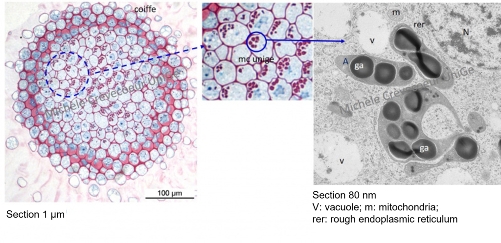
First illustration: cross sections (1μm) through a Zea mays root tip
Below on the left: cross section (1μm thick) made with a diamond knife histo (Diatome) and stained with methylene blue – basic fuchsin. Cell walls and starch grains are stained pink (see detail in the middle), cytoplasm and the nuclei are stained blue. Observation and Imaging : Nikon Eclipse 80i microscope. We then select a part of sample for realization of ultrathin sections (80 nm thick). In this example ultrathin sections have been stained with PATAg (see picture on the right) to reveal starch at the ultrastructural level (figure on the right). The starch grains appear as electron dense globules (ga) and are contained in amyloplast (A).
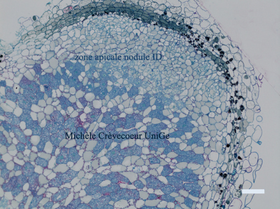
Second illustration : sections through apical part of a ID nodule induced by NGR 234 on Leucaena leucocephala root
On the left parts of longitudinal sections (1 μm; Histo Diatome) through the apical part of an indeterminate nodule (ID) embedded in Epon. Sections have been stained with toluidine blue then observed and imaged with a Nikon Eclipse 80i microscope. Ci : infection thread.
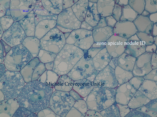
Higher magnification of the meristematic region. ci : infection thread. Bacteria are apparent in the cells located between the meristem (upper part of the picture).
Electron micrograph showing part of a cell in the apical region (meristem) of the nodule, with a polylobed nucleus (N) and small vacuoles both characteristics of a meristematic cell. en: nuclear envelope