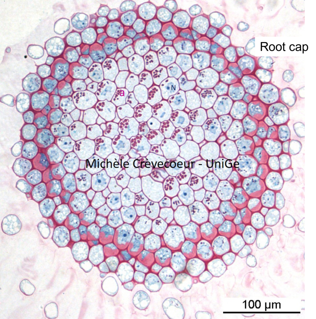
Semithin Epon sections staining with Methylene Blue – Basic Fuchsine
Sections in samples embedded in Epon or LR white are sometimes pre – required and examined with a light microscope before realization of ultrathin sections. Different protocols of stainings are uded to visualize them among which « methylen blue – basic fuchsin (Staining protocol BM BF). Cellulosic cell walls are stained red to pink, the cytoplasm , the nucleus and some cellular organelles are stained blue. Below: illustration of this staining applied to a semi thin Epon cross section in a primary Zea mays root. The section has been made at level of the root cap. In the columella starch grains contained in amyloplasts appear as red – pink granules.