Localization of polysaccharides at the ultrastructural level (Transmission Electron Microscope)
with the PATAg method
Principle of PATAg reaction – Periodic Acid Thiocarbohydrazide Ag proteinate
The reaction is derived from the PAS staining used in classical histology. It is carried out in 3 main steps. First, a treatment with periodic acid specifically oxidizes vic-glycols(vic for vicinity; glycol for free alcohol group on sugar) into aldehydes. The second step consists in treatment of sections with thiocarbohydrazide (TCH) that condenses on aldehydes groups. Finally TCH was made visible as electron dense particles with silver proteinate.
Protocol
The procedure described here below has been applied to ultrathin sections (80 nm thick) made through organs classically fixed with glutaraldehyde – OsO4 and emdedded in Epon. Sections are collected on gold grids covered with a formvar film. All the steps occur at room temperature.
- Float section at the surface of 1% periodic acid drops (~ 30 µl) for 20 min at room temperature. Control grids: deposited on drops of 5% H2O2 instead of periodic acid
- Washes 4 x 5 min on drops (~ 50 µl) of distilled water.
- Float the sections on drops (50 µl) of 0,2 % TCH (Thiocarbohydrazide) in a humid chamber at darkness for 30 min to 72h depending on the material.
- Wash on drops of acetic acid of decreasing concentrations: 20 % (4 x 5 min),10% (5 min),5% 5 min, 2% 5 min et 1% 5 min).
- Wash with filtered (Millipore) milliQ water 5 x 5 min
- Float the sections on drop of aqueous 1% silver – proteinate for 30 min at room temperature at darkness.
- Wash with water 4 x 5 min
- The sections are dried then observed with a Transmission Electron Microscope operating at 80 kV.
Electron microcopes : Philips EM 410; FEI TECNAI TM G2 Sphera
The response e.g. dense deposit of silver grains is positive for many kinds of carbohydrates.
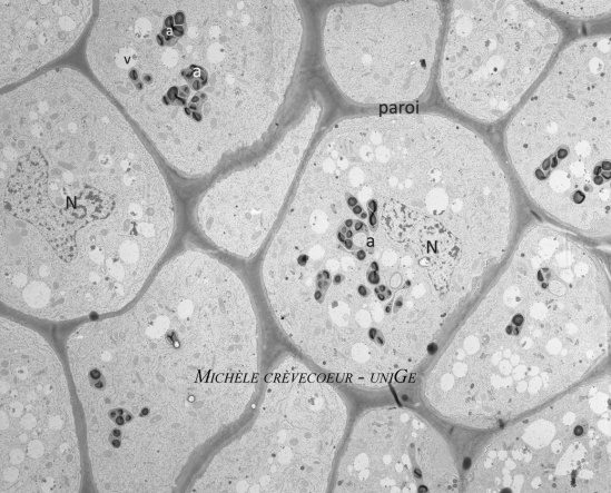
Below parts of ultrathin sections (80 nm) made through a primary root of Zea mays and stained with PATAg.
The first micrograph shows a few cells with postive reaction of starch in amyloplasts (a) and of cell walls that both appear electron dense. N. nucleus
 .
.
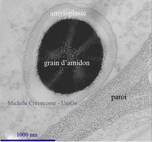
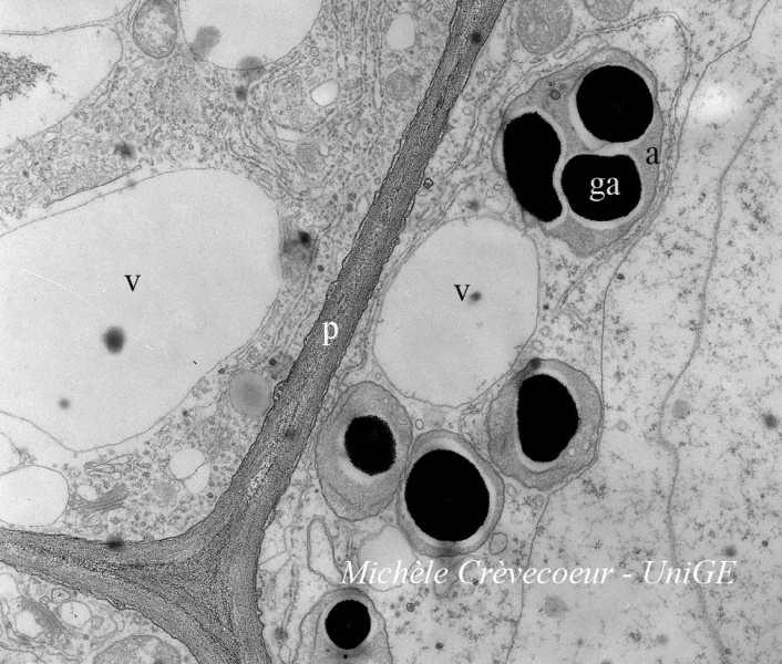
Below two micrographs with details of positive PATAg reaction in Zea mays root cells
On the right detail of a cell wall separating (p) two cells and of starch grains (ga). On the left high magnification view of a starch grain and of the cell wall (paroi) in wich dense deposits of silver appear as small particles.