They are regions of primary growth of roots and stems and of formation of primary tissues in these organs. They are located at extremities of stem and root and are called “apical meristems”, root apical meristem and shoot apical meristem.
Stem cells in primary meristems divide to produce (1) identical cells that will remain stem cells and (2) cells that will differentiate. Mitosis are observed in sections through primary meristems after appropriated staining of paraffin sections. Ex. Feulgen, DAPI,hematoxylin In a young plant the most active primary meristems are those of shoot and root tips. These groups of meristematic cells are already present in the embryoin mature seed, a miniature plant. Three meristematic tissues are observed in these primary apical meristems, in both shoot and root : protodermis, procambium and ground meristem meristem which form primary tissues.
Whereas the root apical meristem is histogen the apical meristem present in shoot tip or in bud has a dual role: it forms tissues (histogen function) and it generates above-ground aerial organs , buds, leaves….(organogenesis function) throughout the lifespan of the higher plants. Shoot branches are derived from axillary meristems initiated in the axils of leaves with the same organization. These axillary meristems function as shoot apical meristem and show the same organization.
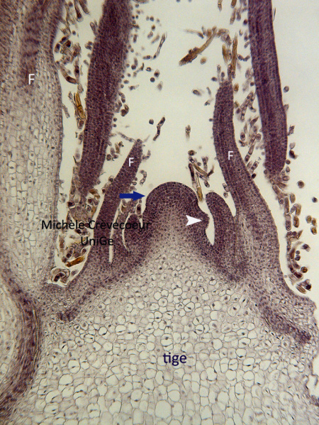
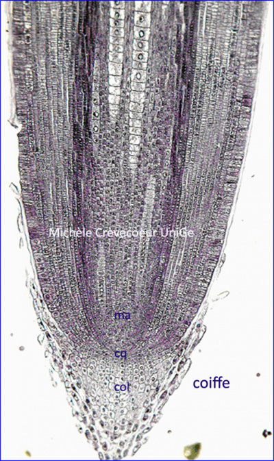
Below microphotograph (light microscopy) of longitudinal paraffin sections in a primary shoot meristem (Spinacia oleracea) on the left and in a primary root on the right (Zea mays) à droite. Sections have been stained with hematoxylin to reveal mitosis.
Cells of primary meristems show typical cellular characteristics easily observable by light microscopy and bytransmission electron microscopy.
F: leaf; blue arrow : apical meristem; white arrow: leaf primordium