DNA staining of paraffin sections with Feulgen method
The Feulgen method is a staining nucleic acid and it is specific of DNA in controlled conditions. It is the most frequent cytochemical method for DNA staining. In addition, it is a method of choice for quantification of DNA in different histological and cytological samples: sections, smears, isolated nuclei. The DNA content is essential for diagnosis and prognosis of tumors and for distribution of nuclei in the different phases of cellular cycle.
The different steps for Feulgen reaction on paraffin sections.
- Deparaffinize
- Hydrolyze in a HCl N solution at 60°C. The length of hydrolysis must be determined for each fixative and each tissue. A control section without HCl treatment is immersed in water at 60°C.
- Wash in tap water for 10 min then in distilled water
- Stain with Schiff’s reagent at darkness and free of oxygen. The time of staining varies according to the tissue and the fixative. For example: 16 min for Zea mays sections through roots fixed with FAA and hydrolyzed 12 min.
- Sulfurous water to remove excess of unfixed Schiff’s reagent.
- Rince in distilled water.
- Facultative step: treatment of sections for 5 to 10 min in 2% sodium metabisulfite (NaHSO3) to be sure that unbound Schiff reagent will not transform in stained fuchsin.
- Dehydrate, clear in xylene and mount in Eukitt, Histokitt or another appropriate mounting medium.
Principle of Feulgen
The staining of DNA using Feulgen method, like the PAS reaction for polysaccharides, is based on the Schiff’s reaction for detecting aldehydes. A pretreatment with HCl N at 60°C for 10 min at room temperature is required. This hydrolysis removes purine bases from the deoxyribose sugar of DNA, unmasking free aldehyde groups. The free aldehyde groups then react with the colorless Schiff reagent which results in purple – magenta staining proportional to DNA content. RNA is not hydrolyzed by the HCl treatment, and the reaction is DNA specific. The intensity of hydrolysis is the critical step: too little hydrolysis and the DNA will be poorly stained; too long hydrolysis and aldehydes will form in the cytoplasm resulting in no specific staining.
The Feulgen staining method has been used to quantify DNA content by micro densitometry /cytophotometry or image analysis.
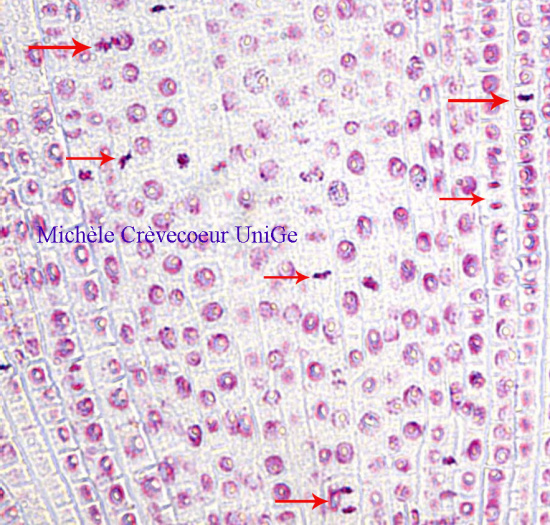
Ilustration of DNA staining with Feulgen in the meristem of a primary Zea mays root.
Paraffin longitudinal section (8 μm thick; red arrows: mitosis).
 f
f