Cross section through a root of Iris
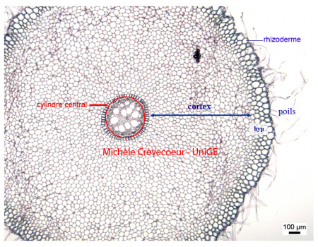
The cross section has been made at the level of root hairs, outgrowths of epidermis. This zone of the root is important for water and minerals absorption. Below the uniseriate epidermis there are 2 to 3 layers of cells (hyp) corresponding to hypodermis.
The root section is divided in two distinct regions, characteristics of roots: a well-developed cortex, constituted of parenchyma cells and a small central cylinder with parenchyma cells in the medium (p).
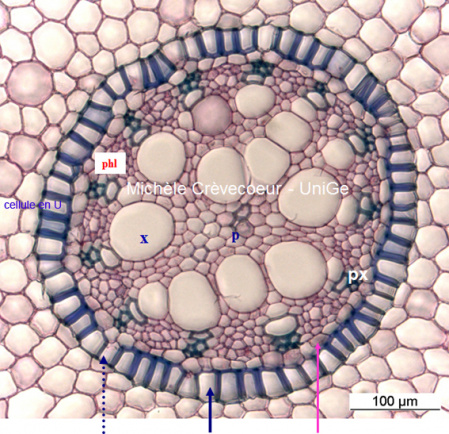
Detailed view of the central cylinder called vascular cylinder. It contains xylem and phloem showing alternating arrangement. Protoxylem poles are in contact with pericycle and metaxylem is towards the center. The phloem consists in patches of cells with thin walls between protoxylem.
The endodermis, the innermost layer of the cortex is single layered and is characterized by striking thickenings of radial and inner tangential walls, with lignin and suberin. A few cells without wall thickening are called passage cells (blue dotted arrows). Just after endodermis there is the pericycle (pink arrow) outer boundary of the vascular (see details on this page).
This cross section illustrates the anatomical characteristics of a young monocot root with two differences with a dicot root (Ex. Caltha palustris): a higher number of xylem and phloem poles and an endoderm with U cells and passage cells.