Trichomes on surface of aerial plant organs.
1/ On the surface of a young bean stem.
Fresh sections made using a vibratome through a young stem of Phaseolus, stained with green methyl and mounted with a coverslip to be observed with a light microscope. Two types of trichomes are observed: glandular and protector.
Below. On the left part of the cross section with vascular bundles (fc) on a concentric row, a characteristic of a young dicotyledon stem. pc: cortical parenchyma; pm: medullar parenchyma. On the right: details of the two types of trichomes: (1) short and glandular and (2) long with sharp tip.
![](https://michelecrevecoeur.ch/wp-content/uploads/2021/12/trichomes-1.jpg)
![](https://michelecrevecoeur.ch/wp-content/uploads/2022/01/Trichomes-3-483x1024.jpg)
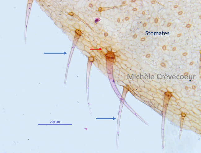
2/ Second example : trichomes on the surface of a pelargonium leaf.
Surface view of the lower face showing random distribution of stomata and trichomes. Blue arrows : non glandular trichomes with basal cells (red arrow).
Mounted preparation: Georges Roux collection. Staining: triple of Flemming.