Micrographs of chloroplasts in Arabidopsis thaliana leaves
collaboration project with the professors Jean David Rochaix and Michel Goldschmidt

 Light micrograph of a cross section in an Arabidopsis th. leaf

Cross section 1 μm thick in a leaf embedded in Epon, made using a diamand knife Histo and stained with toluidin blue.

pp : palissade parenchyma
pl : spongy parenchyma
ep. epidermis
Scale : 100 μm
Comment:  on this micrograph the adaxial face should have been reversed.

Transmission Electron Microgrpah of a cell in the mesophyll.

bottom left (squared) : a detail from thylakoids stacked in grana

A Chloroplast at higher magnification with strach grains (a); m : mitochondria

Chloroplaste de feuille d'Arabidopsis

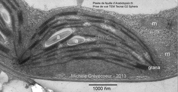
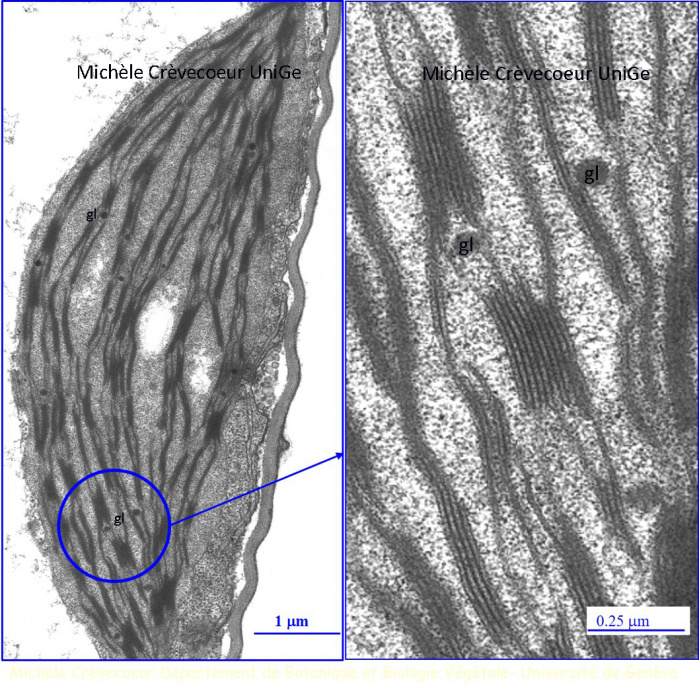
Transmission Electron Micrographs of another chloroplast with details of grana and lipidic globules (g) on the right.

Chloroplaste et grana - feuille d'Arabidopsis