Visualization of stomata at leaf surface in monocotyledons & dicotyledons.
The term stomata design stomatal complexes consisting of kidney or specialized epidermal cells called « guard cells » and the opening (pore) between them. This latter may be closed or open to assure gas exchanges between inside of the leaf and the environment. In some plants two supplementary cells bordering the guard cells, are present and they are called accessory or subsidiary cells.
Stomata can be visualized on sections through leaves and stems. On the other hand, at leaf level they are also easily visualized by peeling epidermis and staining it in vivo. By this way it is possible to get a fast and nice, useful image of stomata and their distribution. Two examples are shown on this page: stomata in bean and mays leaf, a dicotyledon and a monocotyledon.
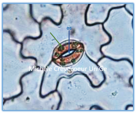
Below: portion of the surface of a Phaseolus vulgaris leaf. On the left random stomata distribution and on the right guard cells with stoma showing a thicker wall in front of stoma. Green arrows: chloroplasts in guard cells, absent in other cells of epidermis.
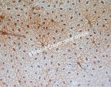
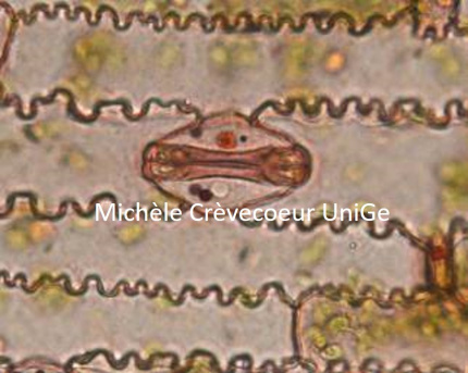
Below: gramineous stomata in Zea mays. On the left distribution in parallel rows; on the right detail of a stomata with two guard cells in form of dumbbell and two subsidiary cells.