Visualization of secretory cells in a stem of rosa.
Micrographs of secretory cells and the protocol to visualize them are shown on this page.
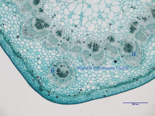
Protocol for realization and staining of sections. Cross sections as thin as possible are made with a soft razor blade on a blade of paper and directly transferred with a paintbrush in a watch glass filled with water. Then the sections are treated with 2,5 % sodium hypochlorite for 10 min. They are washed three times in water and stained with 1% methyl green for 1 to 2 min. The sections are washed again in water, covered with a coverslip and observed with a light microscope. Micrographs of a cross section are shown below
Part of a cross section showing secretory cells scatered in parenchyma, cortical and medullar. They are also present between vascular bundles (fc). x: xylem.
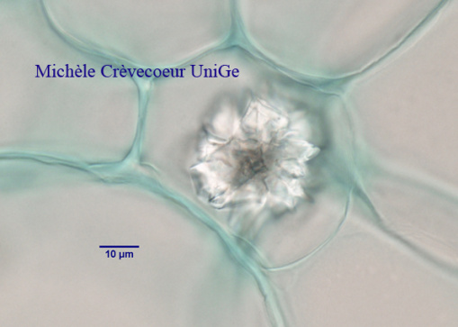
High magnification view of a secretory cell in cortex.