Calcofluor White staining
Calcofluor White (Fluorescent Brightner 28) is a fluorescent blue dye that binds to polysaccharides. This reagent can bind via hydrogen bonds to β (1→ 4) et β (1→3) polysaccharides. It shows a strong affinity for cellulose forming hydrogen bonds with free hydroxyls groups. It is useful to see all cell walls in plant materials using fluorescence microscopy with UV excitation at 365 nm and emission at ~ 440 nm. In medicine it is used for detection of fungal pathogens as it binds to chitin.
Protocol for paraffin sections.
- The protocol is appropriated for plant organs fixed with FAA or aldehydes and embedded in paraffin.
- Deparaffinize and hydrate to water
- Use a pen to draw a hydrophobic circle around sections.
- Carefully remove the excess of water and add a drop (100 µl) of aqueous solution of Calcofluor (Sigma F3543) at a concentration varying from 0.01 to 0,005%. Control of autofluorescence: sections with a drop of distilled water.
- Stain at darkness for 30 sec to 15 min. The concentration and length of staining varies according to the material and the fixative. Examples: for Zea mays roots fixed with FAA 30 sec in 0,01%; for Quercus roots fixed with formaldehyde – glutaraldehyde, 5 min with 0,1%.
- Wash slides thoroughly with distilled water with gentle agitation.
- Mount in appropriating medium such as Vectashied. (Antifading mounting medium)
- Observation: fluorescence microscope with appropriated filters. For Leica DM IRE 2: A filter for excitation (UV: 340 – 380 nm); For Confocal Leica SP2: excitation with laser 405 nm; emission: 450 to 547 nm.
A stock solution of calcofluor can be prepared and stored at darkness at 4°C for months.
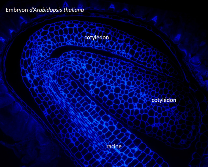
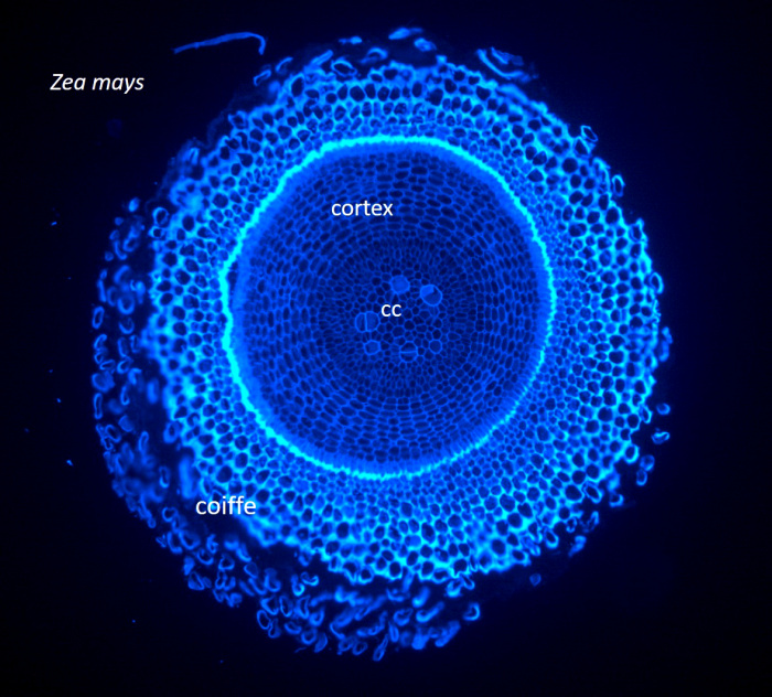
Two micrographs of paraffin sections stained with calcofluor white are shown below. They were observed and imaged with a Leica DM IRE2 microscope (excitation at 340-380 nm; emission at 425 nm) equipped with a Leica DC300F camera.
The fisrt one is a cross section through a Zea mays primary root with root cap at the outside. Cellulosic cell walls are all fluorescent and there is autofluorescence of lignin and suberin. cc : central cylinder.
Microphotograph of a paraffin section through an Arabidopsis thaliana embryo