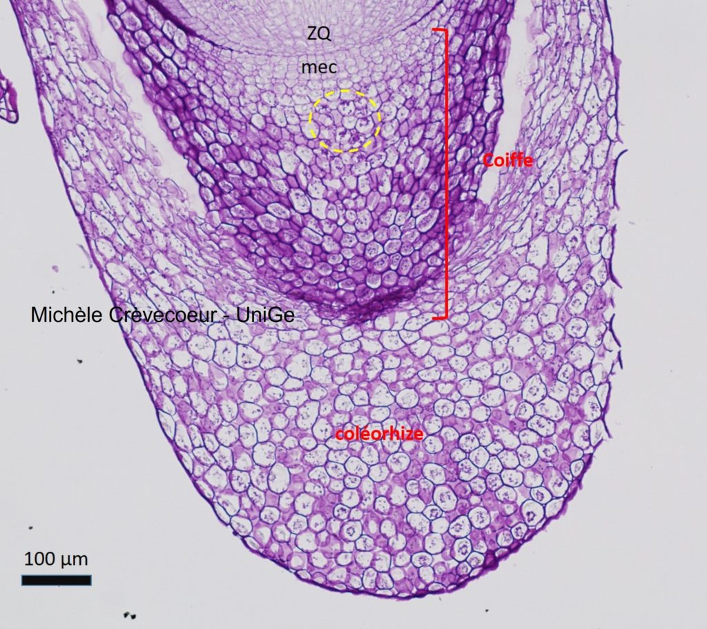
1/ Micrograph of a longitudinal section. This is a paraffin section (10 μm) through a Zea mays embryo fixed with FAA. It was stained with PASto visualize polysaccharides. The root cap at the extremity is protected by a protective tissue called « coleorhiza« . The root cap and the coleorhiza are both constituted of parenchyma cells with a thin cellulosic wall.
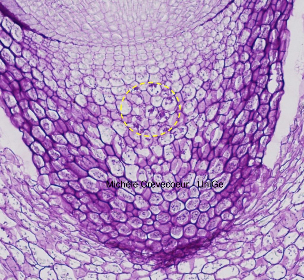
mr : root meristem; cq : quiescent center; mec: méristème d'entretien de la coiffe. On the right higher magnification of the root cap.
In the yellow dotted circle in the root cap, we distinguish small grains that are pink fuchsia stained, They correspond to starch grains that are involved in gravity perception by the root (positive gravitropism) and are contained in amyloplasts.
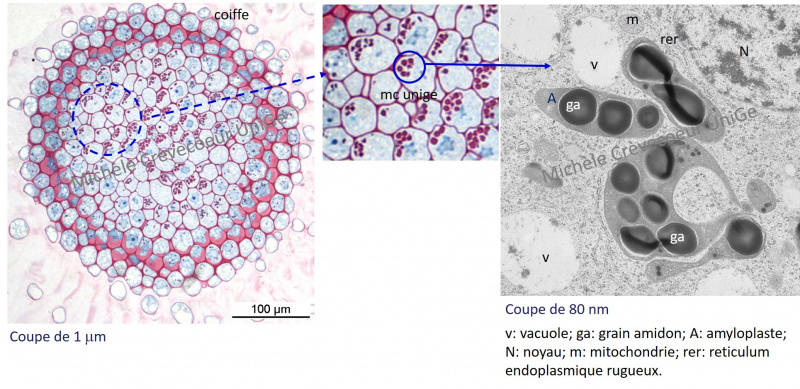
2/ Micrographs of a cross section through the root cap. Epon cross section (1μm) made with a diamond knife Histo and stained with methylene-blue basic fuchsin. Starch grains appear as pink stained globules (detail in the middle). On the right, electron micrograph showing four amyloplasts (A) containing each several starch grains (ga) in a root cap cell. They appear as electron dense.