Starch Storage Parenchyma
Two Protocols of starch localization are given below and illustrated by micrographs
First example: staining of starch in potato chair using fresh sections and Lugol reagent.
Starch is an energy reserve for potato and consists of long chains of glucose molecules. It contains a mixture of amylose and amylopectin and is present in the form of granules varying in size from 1 to ~ 100 µm. Inside the granule the starch molecules are arranged into concentric layers around a central point called hilum, starting point for the accumulation of amylose and amylopectin
Material required
Watch glasses
Thin Razor blade
Brush to collect sections.
Lugol: Iodine/Potassium iodide solution
Preparation of Lugol
2% Potassium iodide (KI) and 0,2 % diiode (I2)
Protocol
Sections as thin as possible are made with a thin and soft razor blade in a piece of potato chair.
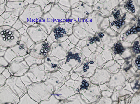
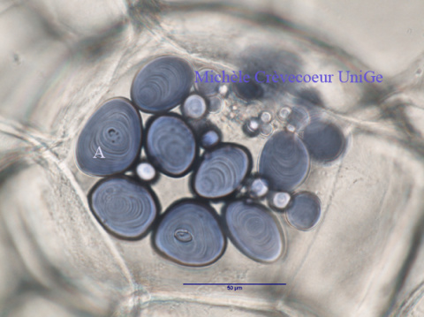
The sections are directly transferred with a paint brush into a watch glass containing water. They are then immersed into a drop of Lugol solution for 1 min and directly collected on a microscope slide in a drop of water. They are covered delicately with a coverslip and observed with a light microscope. Starch grains appear as blue to purple particles.
Micrographs illustrating Lugol staining
On the left parenchyma cells that compose the potato chair with starch grains-stained blue purple. They are contained in unstained amyloplasts and appear as oval structures.
On the right higher magnification picture of starch grains (A). They appear composed of a series of concentric layers.
The starch grains are contained in amyloplasts that are colorless. Hilum is visible in the center of a few strach grains.


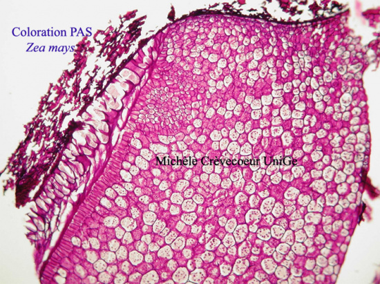
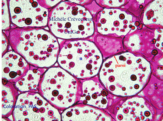
2nd Example: storage starch parenchyma in endosperm of a corn caryopsis, popular name grain, typical dry indehiscent fruit of Poaceae or Gramineae. Endosperm is the tissue surrounding and nourishing the embryo in the seeds of angiosperms.
Part of a paraffin section through a Zea mays endosperm stained with PAS. On the right detail of cells in which starch grains appear as pink globular structures (a) and pink stained cell walls.The nucleus (n) and the cytoplasm are not stained.

