Sclerenchyma : fibers
Lignified fibers (to distinguish from cellulosic fibers) commonly occur in groups forming strands associated to vascular bundles in the stem. They are also found in “thick” leaves. They have a thick secondary lignified wall, and they are dead at maturity without protoplasm.
Two examples are shown on this page: parts of cross sections in a stem of a Viscum and in a leaf of holly
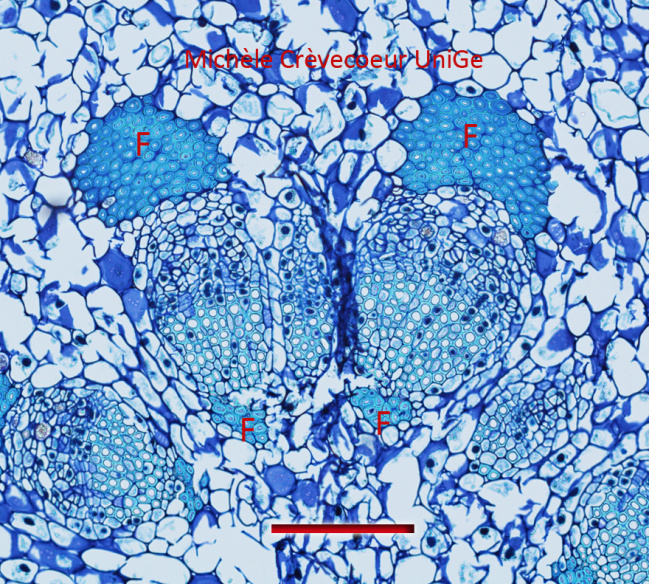
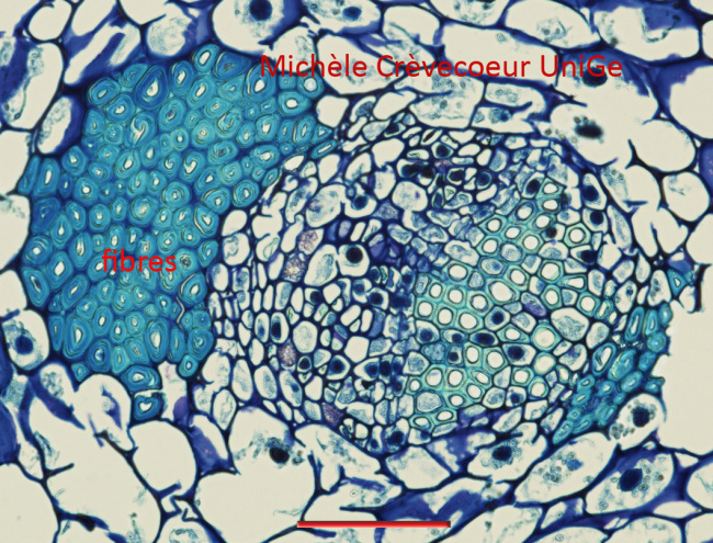
1/ Part of a cross section in a stem of Viscum album (see stem anatomy) stained with toluidine blue. In the first micrograph we observe two vascular bundles with strands of fibres (F) at both extremities. They are less numerous at the extremity located toward the center of the stem. The second micrograph shows the vascular bundle at higher magnification.
Scale: 500 μm; 100 μm (below).
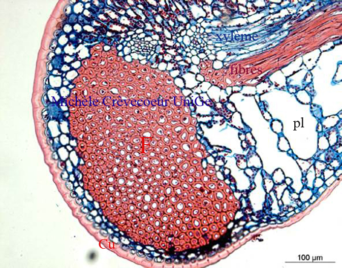
2/ Part of a paraffin cross section through a leaf of Ilex aquifolium stained with safranin – alcian blue (See anatomy of holly leaf) with strands of fibres (F). pl: spongy parenchyma.