Sclerenchyma, supporting lignified tissue: sclereids.

Three examples are shown on this page to illsutrate sclereids: peer chair, root and leaf surface of waterlilly.

1/ Sclereids in the pear fruit.

The gritty texture of pear fruit results from the presence of sclereids. When we eat a pear we have the sensation of small grains that are sclereids.
They are easy to be imaged: a small piece of chair is selected in which we cut thin slices with a razor blade deposited in a watch glass and stained 1 min in a drop of toluidine blue (0,05%). After 2 rinses in water, they are transferred on a slide in a drop of water, covered with a coverslip and directly viewed under a light microscope.
The sclereids are blue stained and appear as stone shaped. They are called stone cells.

Scale : 100 μm
Scale : 50 μm

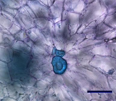
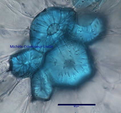
2/ Sclereids in waterlilly root.

Another example of sclereids found in an aquatic plant, water lily. They are present in the different organs (root, stem, leaf and petiole) of this plant living in water that require supporting tissue to allow flat large leaves to float at the water surface. They are called astrosclereids.
They are also illustrated in a cross section trhough a root of this plant (see root anatomy) in which sclereids are observed in different places in the aerenchyma (blue arrows).

Site Nymphea racine

3/ Sclereids at the surface of a leaf of waterlilly.

Sclérites feuilles nénuphar.jpg

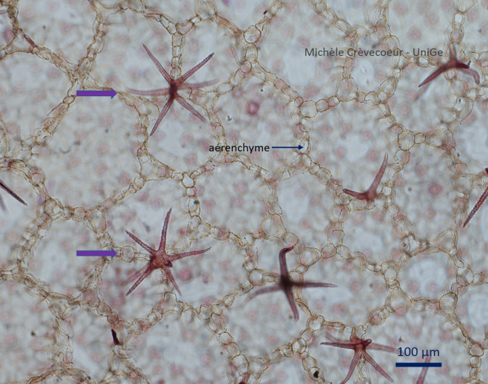
Surface view of a  waterrlilly leaf on which we observe many sclereids in form of star. They are called astrosclereids (violet arrow) and are composed on one cell with several arms. Aerenchyma with small cells disposed around large air spaces (lacunae) is clear.