Collenchyma : cellulosic support tissue of young growing organs
Examples below: light microphotographs of collenchyma with parts of cross sections in a young stem of sage and in a celry petiole to illustrate angular collenchyma.
1/ Angular collenchyma in a young stem of Salvia (Sage)
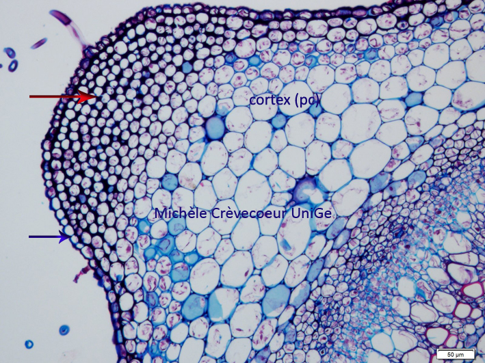
Part of a paraffin cross section stained with toluidine blue. Under the epidermis (blue arrow) several layers of angular collenchyma are observed (red arrow). Intercellular spaces are lacking and filled with cellulose thickenings. After the collenchyma there is the cortical parenchyma (pc) consituted of large cells with thin cellulosic walls and small intercellular spaces.
2/ Angular collenchyma in a celery petiole.
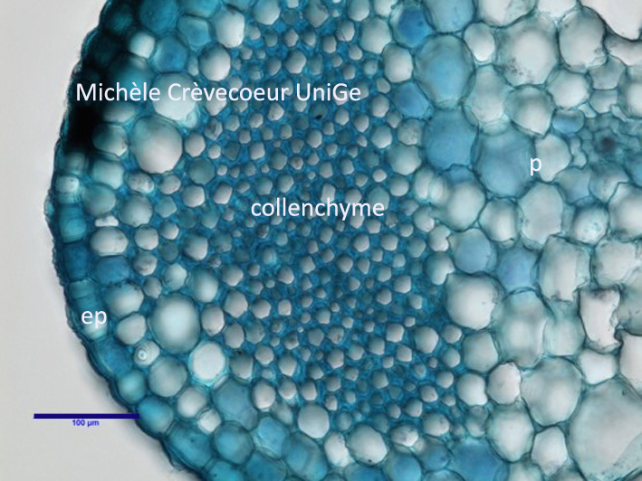
Part of a fresh cross section made with a razor blade and stained with toluidine blue.
p : parenchyma; ep : epidermis
See also: microphotograph of a part of a cross section stained with PAS