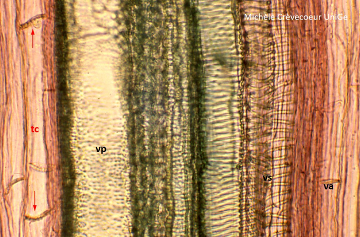
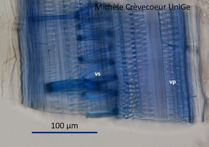
Primary vascular tissues in longitudinal sections Xylem vessels and sieve tubes are easier to be recognized on longitudinal sections than on cross sections. However cross sections give important information ontheir distribution in different organs and allow to determine in which organ a section is made and whether it is a monocotyledon or a dicotyledon. Microphotographs on this page illustrate two portions of longitudinal sections. The first microphotgraph below illustrates part of a longitudinal section at the level of primary xylem and phloem in a young stem of Cucurbita pepo. From the right to the left we find vessels with different lignin thickening of the walls: annular vessel (va), spiral vessel (vs) and punctuated vessel (vp). On the left two phloem sieve tubes (tc) with sieve plates (red arrows). The second microphotograph shows part of a longitudinal section, stained with tlouidine blue, at the level of xylem in a young Zea mays shoot. vs : spiral vessel; vp : punctuated vessel.