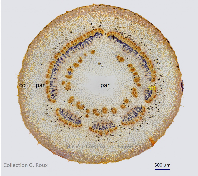
The micrographs on this page illustrate sections from George’s Roux collection. The cross sections have been made in a petiole of Common Fig in Moraceae family. The two first micrographs illustrate a section after Flemming staining. Walls of the different cells are stained yellow, except those of xylem that are stained violet. The vascular bundles are distributed on a circle with xylem toward center and phloem toward outside. Col: collenchyma; par: parenchyma; fc1: a vascular bundle
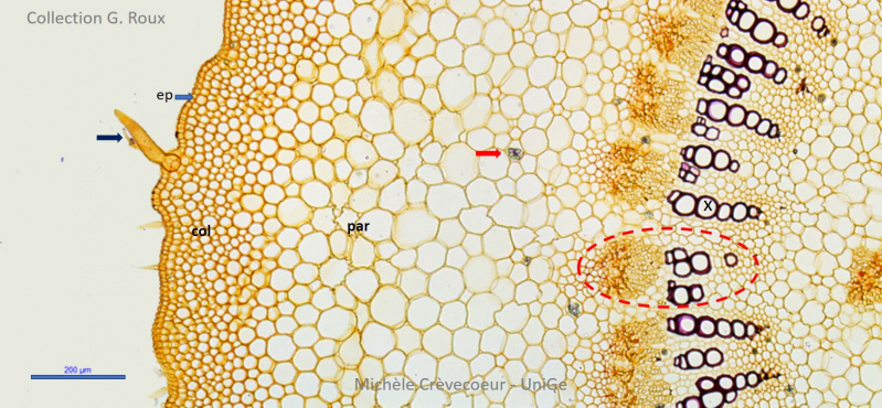
Below part of a cross section with the different tissues from the periphery towards the center : the epidermis (ep) with trichomes (blue arrow), a few layers of collenchyma (col), ground tIssue or parenchyma (par) with aggregates of calcium oxalate crystals in some cells (red arrow), the vascular bundles (dotted red contour) with xylem (x) towards the center of the petiole
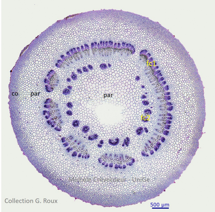
Below: micrograph of a section stained with carmin – green iodide. Walls of the cells are stained blue. co : collenchyma; par : parenchyma; fc1 et fc2 : vascular bundles.