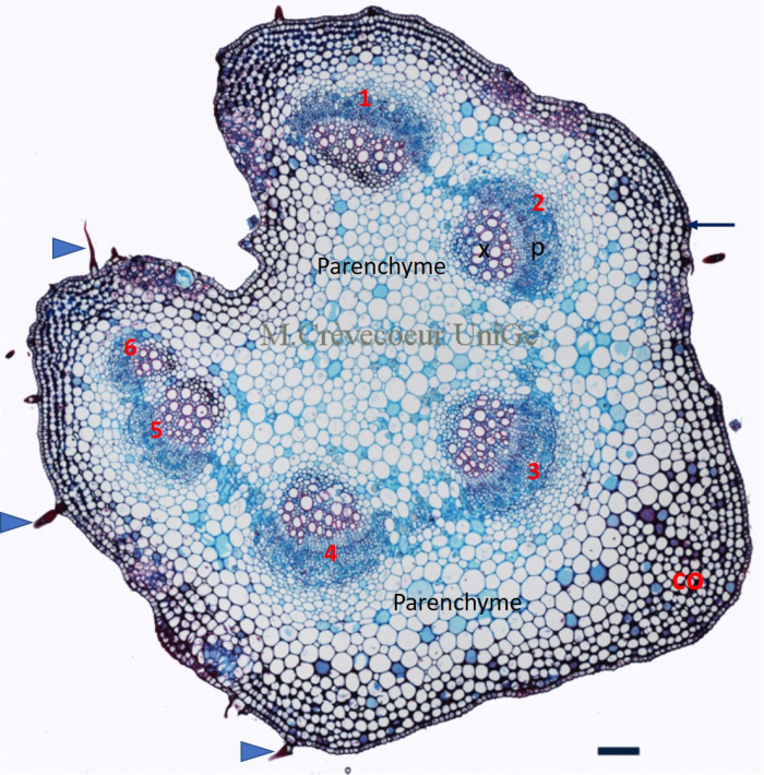
Cross section in a petiole of nettle
It is a paraffin cross section stained with astra blue – basic fuchsin. The blue arrows indicate trichomes that are extension of cells of the uniseriate epidermis. Six vascular bundles are distributed on a ring as in the stem. Parenchyma is present in the center of the organ and between epidermis and vascular bundles. Three to four layers of parenchyma below epidermis consist of support tissues, angular collenchyma (co) on the whole periphery of the petiole.
Scale: 100 μm.
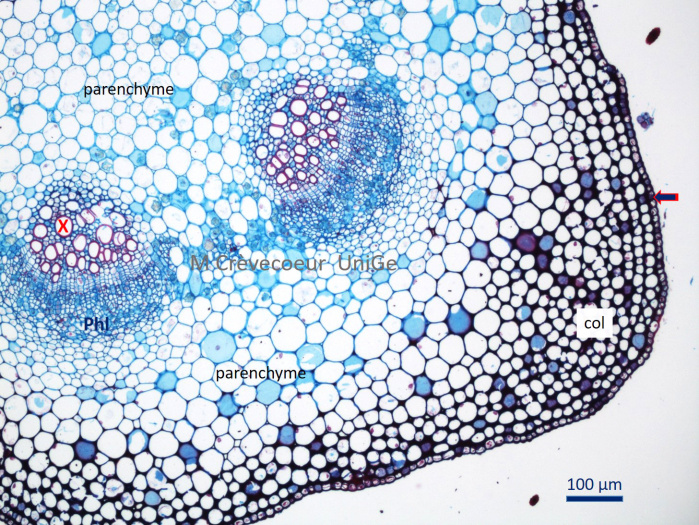
Part of the section showing two vascular bundles with xylem (x) towards the center, phloem towards the periphery (phl) and cambium between the two tissues. Layers of collenchyma (col) support tissue with cellulosic cell walls, are observed below epidermis.