Cross section in a leaf of Quercus – oak
The sections have been made in a portion of leaf fixed with FAA and embedded in paraffin. They have been stained with
safranin and alcian blue.
Micrographs below show tissues at the level of midrib (upper) and at the level of lateral veins (bottom).
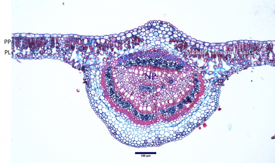 PP: palisade parenchyma ; PL: spongy parenchyma; NP: midrib
The mesophyll is herogeneous with palisade parenchyma towards the adaxial face and spongy parenchyma toward the abaxial face. Stomata are observed in abaxial epidermis and both epidermis have a thin cuticle.
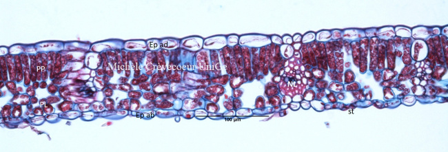 Ep ad: epidermis adaxial face; Ep ab: epidermis abaxial face; st: stomata;
PP: palisade parenchyma; PL : spongy parenchyma; NL: lateral vein with vascular tissues.
Ep ad: epidermis adaxial face; Ep ab: epidermis abaxial face; st: stomata;
PP: palisade parenchyma; PL : spongy parenchyma; NL: lateral vein with vascular tissues.