Cross section in a leaf of Calathea lutea
Calathea lutea, that have many common names among which Cuban Cigar, belongs to Marantaceae family and is native to tropical America.
It is an evergreen, rhizomatous, perennial, plant that can grow up to 2 – 5 meters tall. Calatheas are frequently used as ornamental plant. In natural environment they are found in moist or swampy tropical forests. Calathea lutea is cultivated as an ornamental plant and the leaves are harvested by people for thatching and making woven baskets.
The leaves of Calathea lutea are simple, oval to elliptic and coriaceous with a long petiole (1,2 to 2 m) and large blade (1m long and 40-50 cm wide). They show complex movements involving both elevation and folding of the leaf surface, as in other plants in Marantaceae family known as “prayer plant family”. These movements are possible thanks to the presence of a pulvinus, a specialized sheath at the tip of the petiole. The leaves unfold in the morning and are horizontally oriented and flat to maximize light absorption and they straight up at night, folding in half along the midrib. This daily movement of the leaves, in accordance with a circadian rhythm is known as “nyctinasty”.
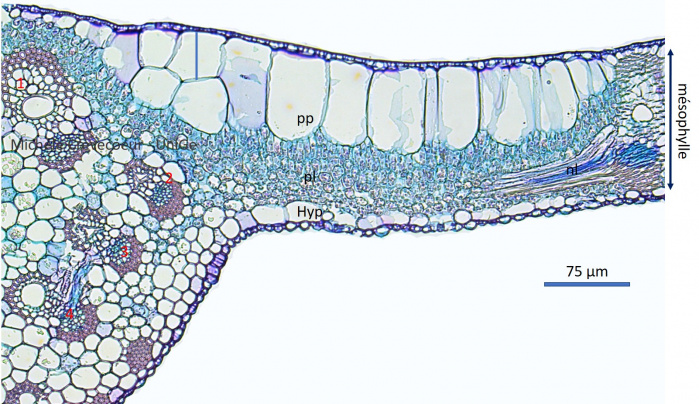
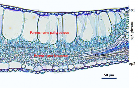
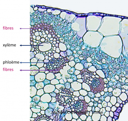
The micrographs below illustrate a cross section (10 μm) in a portion of a leaf fixed with FAA and embedded in paraffin. Sections have been stained with Astra blue and Basic Fuchsin.
Below cross section at the level of the midrib with 4 vascular bundles (numbers 1 to 4) and the lamina on the right. The mesophyll is heterogeneous with palisade parenchyma composed of large cells (pp) toward adaxial face and spongy parenchyma (pl) toward abaxial face. A layer of hypodermis (hyp) is observed on the abaxial face.

Below details of the section showing: (1) on the right, the lamina with the two parenchyma and epidermis of both faces (ep1 and ep2) that show a thin cuticle; (2) on the left, the vascular bundle of the midrib with superposition of xylem (x; towards the adaxial face) and phloem and fibers with thick walls on both sides. A lateral vein is seen in longitudinal section.

