Cross section in a leaf of Urtica dioïca – nettle
Urtica dioica, often known as common nettle, is an herbaceous perennial flowering plant in the family Urticaceae and comes from northern Europe and Asia. The leaves are pointed, with toothed edges. The faces are also covered with fine teeth.
The sections on this page are paraffin sections (8 μm thick) stained with astra blue and basic fuchsin.
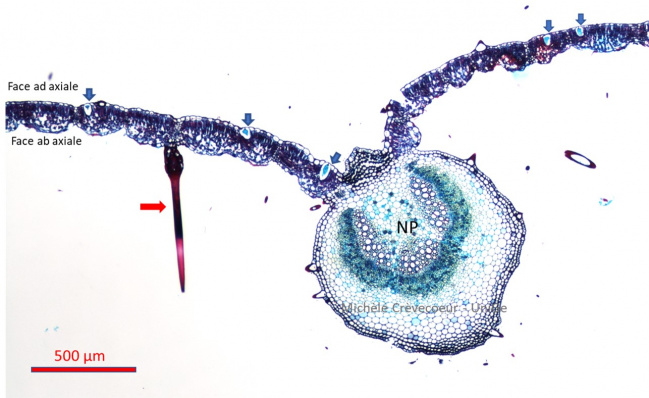
The first micrograph shows the tissues at the level of main vein (NP) with mesophyll cells from either side. Several cystoliths, calcium carbonate deposits, are observed on the adaxial side (5 blue arrows) and a long, pointy trichome (red arrow) on the abaxial side.
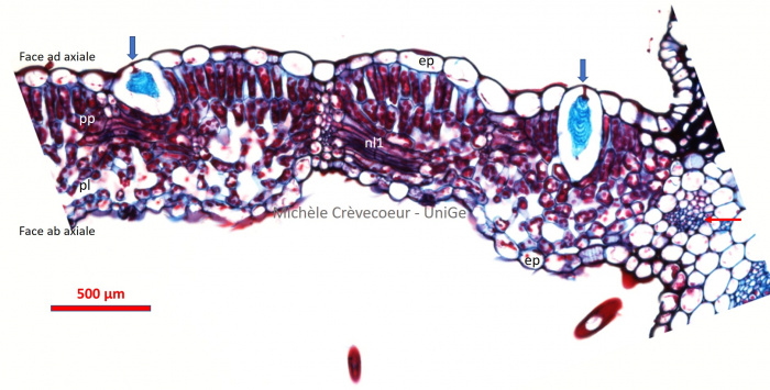
Below : tissues in a portion of the section with lateral veins (nl1 longitudinal view; nl2 cross section; red arrow). Two cells of the epidermis, litocysts (blue arrows), are observed on the adaxial epidermis. They are large and contain cystoliths (blue stained), encrusted onto a stalk of cellulose (in red) connected to the wall of the cell. Between the two epidermis (ep) the mesophyll is heterogeneous, with palisade parenchyma (adaxial face) and spongy parenchyma (abaxial face).
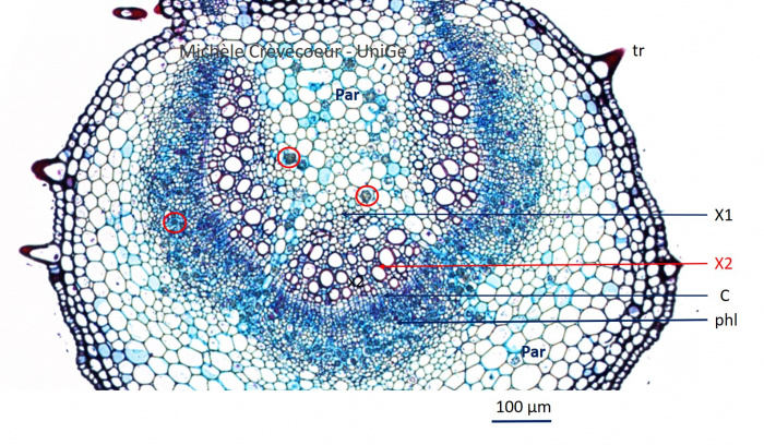
Below : high magnification view of the vascular tissues of the main vein. From the adaxial toward the abaxial face we find successively: xylem 1 (x1), xylem 2 (x2), cambium (c) and phloem. From either side there is parenchyma (par). Numerous aggregates of oxalate crystals are observed in the central parenchyma and in the phloem (red circles).