Zea mays root imaged by Scanning Electron Microscopy
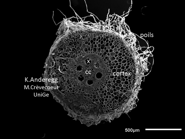
The first micrograph below illustrates a cross section at low magnification. It is characterized by a small central cylinder (cc) and a large, well developped cortex. The section has been made at the level of the piliferous region as indicated by the numerous hairs at the rhizodermis level.
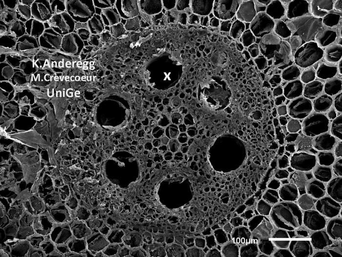
Detail of the central cylinder with the large xylem vessels (X). The endodermis is not well differentiated.
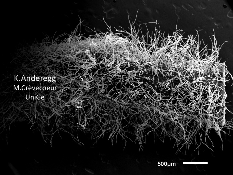
Surface view of the piliferous layer illustrating the very high density of root hairs in this region of the root. It functions in the process of uptake of water and nutrients form the soil.