Triticum durum imaged leaf by Scanning Electron Microscopy
Samples have been collected at the botanical garden at Geneva with collaboration of Dr Fred Stauffer, conservatory
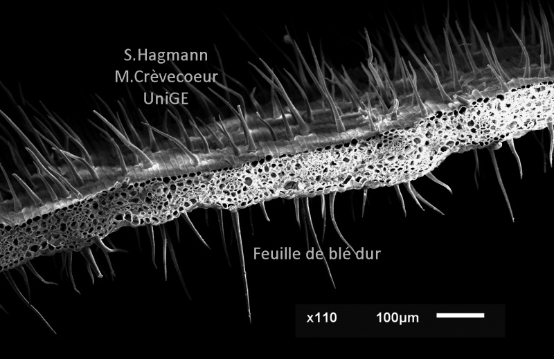
First micrograph : Low magnification view of a wheat leaf in cross section. Trichomes are observed on both faces and the mesophyll between the two epidermis is homogeneous.
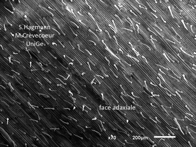
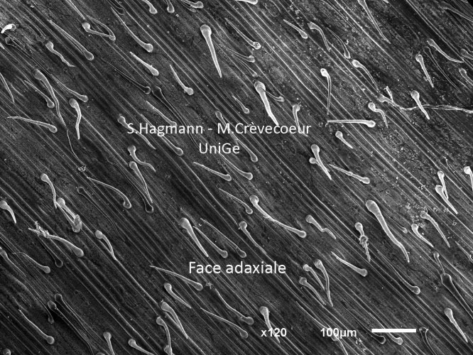
Appearance at low magnification of the adaxial face of a leaf: it is covered with non - glandular trichomes distributed in parallel rows.
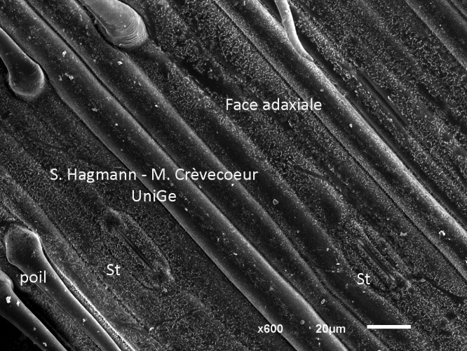
Another appearance of the adaxial face at higher magnification with stomata (st) two on this micrograph. They are also distributed in parallel rows.
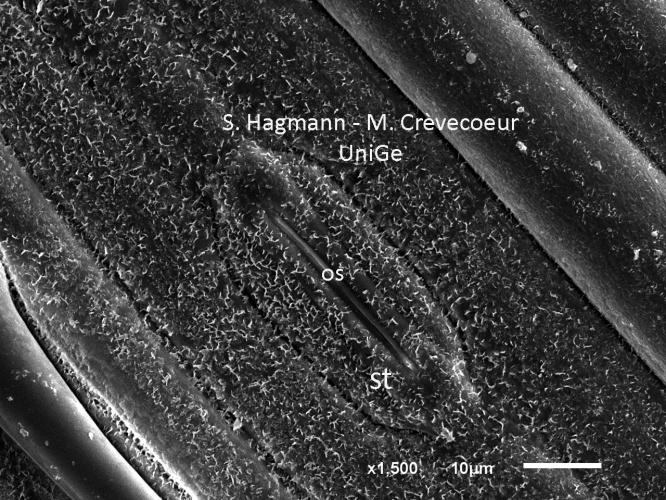
Stoma on the adaxial face at higher magnification (os : ostiole)
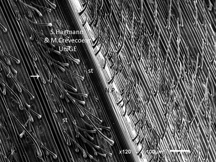
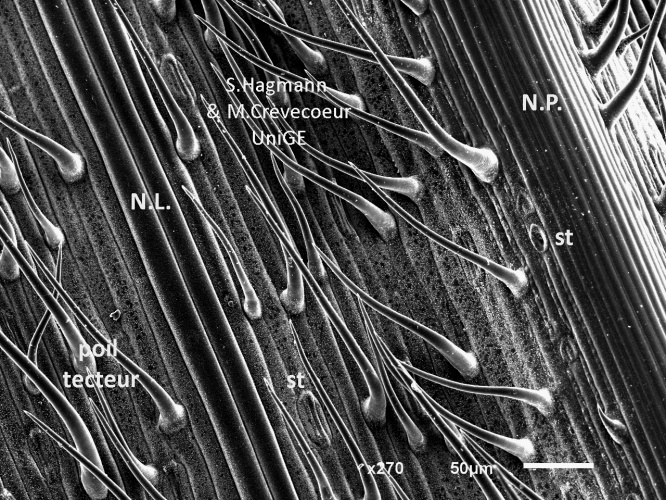
Abaxial face of the leaf with the main vein (NP). The face of the lamina and the main vein are both covered with non - glandular, elongated and tapered trichomes. They are distributed in parallel rows as on the the adaxial face. Stomata (st) are observed apart from the vein.
Appearance of the abaxial face at higher magnification with the main vein (N.P.) and a lateral vein (N.Nl.). Stomata (st) are observed apart from the vein and are distributed in parallel rows as the non - glandular trichomes