The sections are paraffin cross sections (8 μm) stained with safranin and alcian blue. Tissues with cellulosic walls are stained blue and those with lignified walls are stained pink.
Below: two micrographs at the level of the main vein. On the right, higher magnification of its vascular bundle showing characteristics of the Krantz anatomy, typical of C4 plants. Mesophyll cells radiate outwards from the bundle sheath enclosing the veins and containing RUBISCO. As clearly seen at the ultrastructural level (transmission electron microscopy) the chloroplasts of mesophyll cells contain grana whereas they are usually without grana in the bundles – sheath cells.
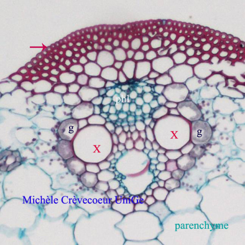
Pink arrow: epidermis and below layers of fibers with thick lignified walls. x: xylem vessels; phl: phloem; g: perivascular gain.
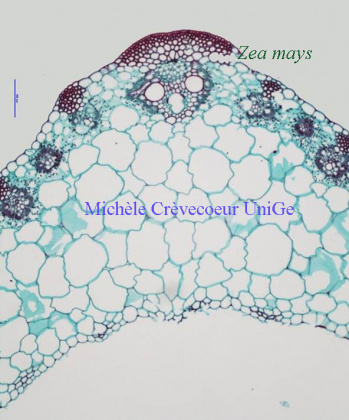
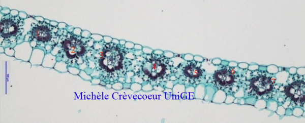
Part of the section in the lamina with secondary veins and their vascular bundles corresponding to veins numbers 1 to 7. The mesophyll is homogeneous. It does not have distinct palisade and spongy parenchyma, a characteristic of leaves with two similar morphological faces. The vascular bundles are distributed on a row and correspond to parallel veins, a characteristic of a monocotyledon.
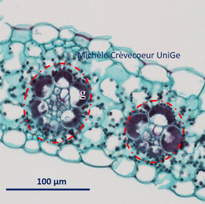
Part of mesophyll with 2 vascular bundles (dotted red circles) and the sheath (g) of cells around them.
Characteristics of this section corresponding: To a monocotyledon: vascular bundles on a row; V distribution of xylem vessels with phloem between arms of this V and homogeneous parenchyma in the mesophyll.
To a C4 plant (Poaceae): bundle sheath cells around each vascular bundle. The chloroplasts in the sheath and the mesophyll are organized to allow carbon dioxide to concentrate in bundle sheath around RUBISCO.