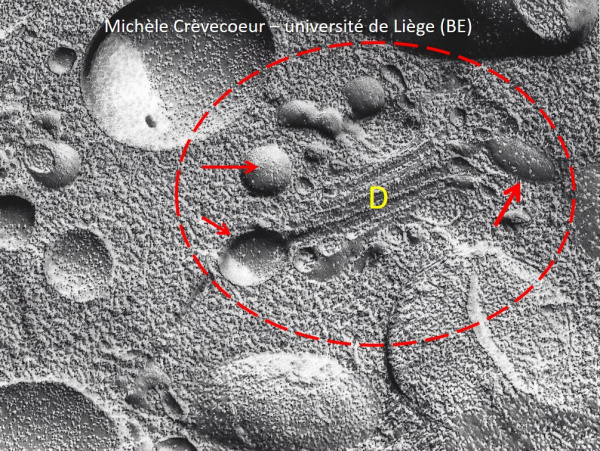
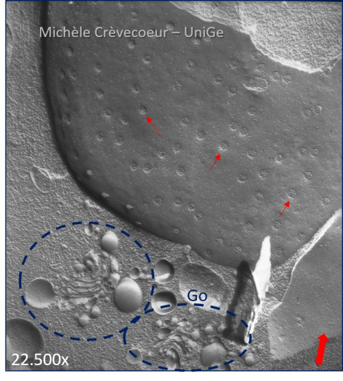
Golgi elements (dictyosomes and vesicles) imaged on cryofracture in mays and spinach cells.
On the left transmission electron micrograph of a Golgi element in a Zea mays root cell. It consists of a stack of sacules forming a dictyosome (D) with vesicles at both extrmities (red arrows) whose surface is covered with proteins. Gx: 60.000.
On the right part of a replica in a spinach shoot meristem. It illustrates the proximity of the nucleus and the Golgi with two elements (blue dotted circles) constituted of dictysomes and vesicles. The small red arrows indicate nuclear pores.
Direction of shadowing is indicated by the red arrow at the bottom right