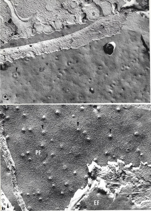
Fracture faces of plasma membrane In the primary root of a Zea mays embryo germinated 24 h.
Above: extracellular (EF) and protoplasmic (PF) faces of the plasma membrane. On each face we observe cellulose microfibrills of the wall (P) between two adjacent cells. The density of intramembrane proteins is higher on protoplasmic faces. The arrows indicate the plasmodesmas that appear as protuberances on the PF face and as holes on Ef face
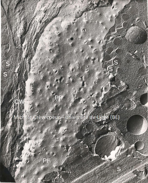
Above: From the left we successively find : spherosomes of a cell (S), cellulose microfibrills of the wall (CW) between two adjacent cells and protoplasmic face (PF) of the plasma membrane. Gx: 17.650.