Cross section through a stem of Urtica
The nettles are a genus from Urticaceae family which includes about thirty species of herbaceous plants with hairy leaves and a quadrangular stem. Micrographs below illustrate a paraffin cross section (10 µm thick) through a portion of stem fixed with FAA and stained with Astra blue – basic fuchsin.
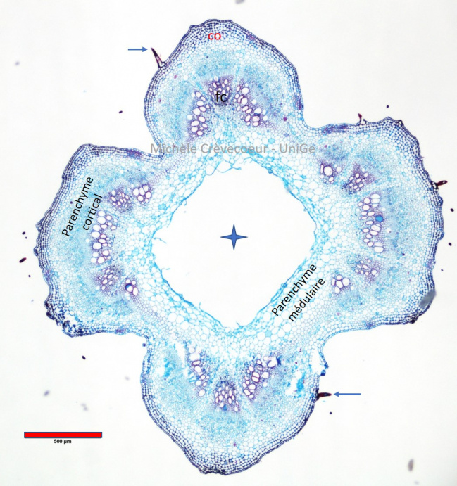
General aspect of the section. Central part of the pith has disappeared and appears as a large lacuna (star) and the stem is hollow. The vascular tissues (fc) are distributed on a single ring at the periphery of the pith, with xylem and phloem superposed. Xylem vessels are located towards the center and the phloem towards outside (characteristics of a young dicotyledon stem). Trichomes are observed at the epidermis level (blue arrow).
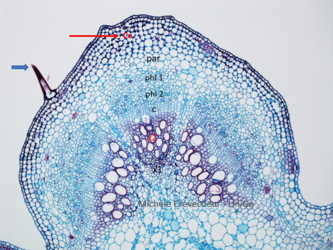
Part of the section. From the center towards outside we successively find: primary xylem (x1); secondary xylem (x2), a few layers of rectangular cells corresponding to cambium ( c), secondary and primary phloem (phl2 and phl1, cortical parenchyma (par); the angular collenchyma (co) constituted of thick – walled cells and the epidermis.
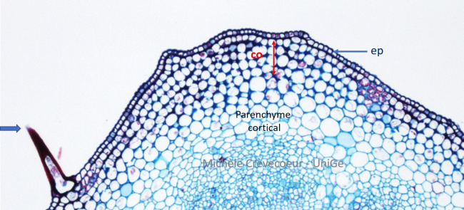
Portion of periphery of the stem section: uniseriate epidermis (ep) with trichomes (blue arrow on the left), a few rows of collenchyma (co) with thick walls very apparent in corners of cells and the cortical parenchyma with small intercellular spaces.