Microphotographs of a cross section through a stem of Salvia officinalis – Officinal sage
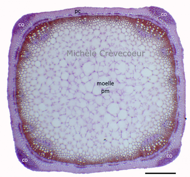
It is a slide from the collection of Mr Georges Roux. The first micrograph shows the general aspect of the section with an extensive central pith (pm: medullar parenchyma) composed of large polygonal cells with a thin cellulosic cell wall and a cortex reduced to a few rows of parenchyma (pc) cells. The section has a square shape with clusters of collenchyma (co) in the four corners of the section. Scale : 500 μm.
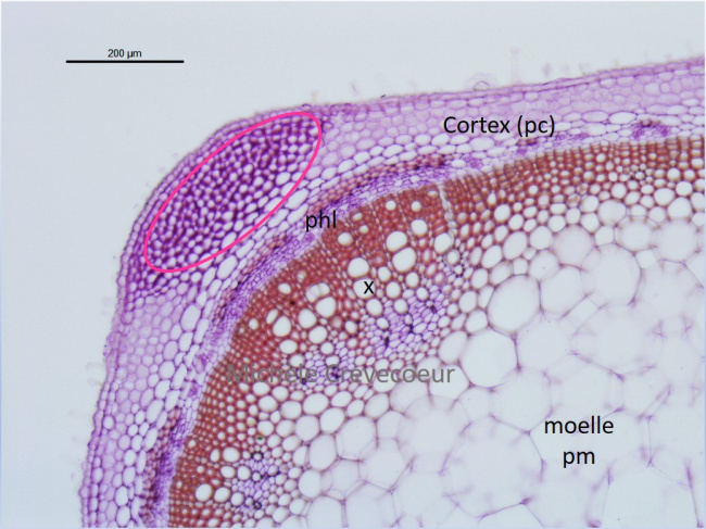
Portion of the section at higher magnification with the angular collenchyma (oval pink) and the superposed xylem (X) and phloem (phl) with the xylem towards the center of the stem.