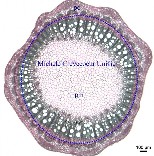
Microphotographs of a cross section through a two years old stem of Vitis Vinifera
This cross section perfectly illustrates the secondary growth with the presence of secondary vascular tissues, secondary xylem and secondary phloem, in a two year old stem of vine. The cellulosic tissues are stained pink and the tissues with lignified cell walls are stained green.
The central part of the section consists in a well-developed pith (medullar parenchyma ; pm) constituted of large polygonal thin – walled cells and contains vascular tissues. The cortex is reduced to a few layers of cells between epidermis and fibers associated to vascular bundles which are distributed on a single ring (bleu dotted ring) a characteristic of dicotyledon stem.
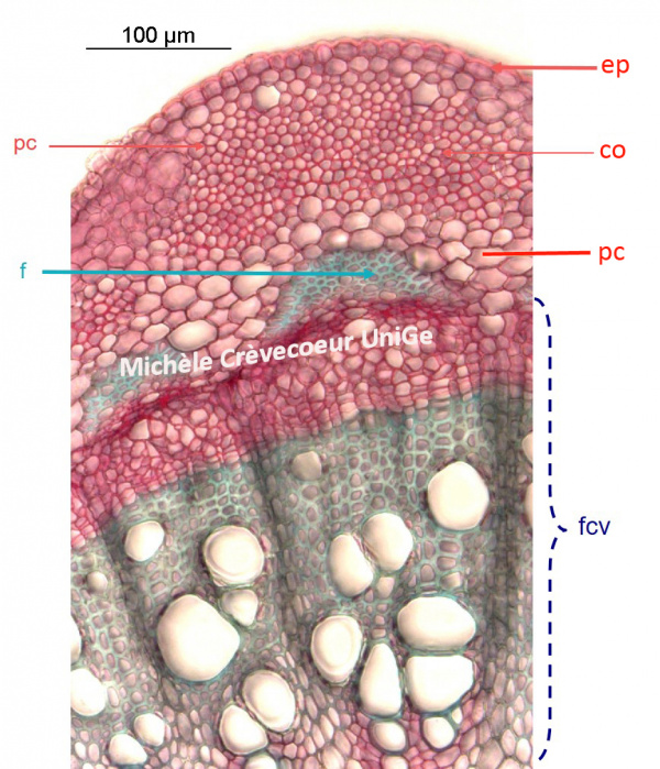
Below, part of the section. From the outside to the center we successively find: epidermis (ep) a monolayer of thickly cells, the cortex (pc) composed of a few layers of parenchymatic cells, the collenchyma (co) with thick cellulosic cell walls, a few larger cells of cortical parenchyma (pc), fibers (in green) with lignified thick cell walls and a vascular bundle (fcv).
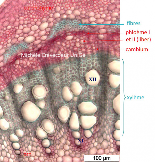
Here below part of the section with high magnifications of secondary vascular tissues, xylem II and phloem II. Both tissues are superposed with xylem towards the center and phloem towards the periphery, a common characteristic of a dicotyledon stem.
Layers of cambium, the secondary meristematic tissue forming secondary xylem and phloem, are well apparent.
As the result of secondary growth, the primary phloem of the vascular bundle has been displaced outwards and due to cellulosic walls of its cells it appears compressed against the fibers. The primary xylem has been displaced towards the center.
XI : primary xylem; XII: secondary xylem