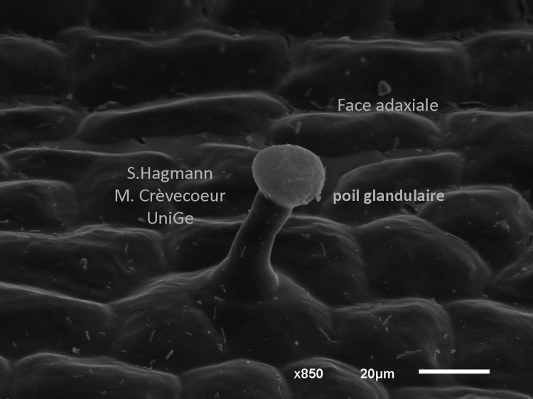
Scanning electron micrographs of Rosmarinus officinalis (common rosemary) leaf Samples collected at the Geneva Botanical Garden with collaboration of Dr Fred Stauffer, Curator.
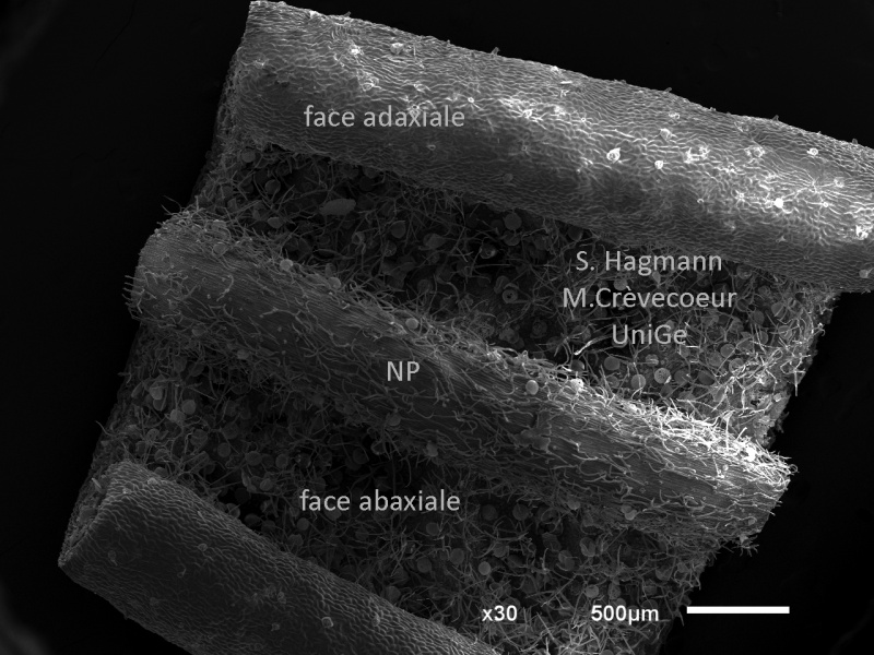
View of a part of a leaf at low magnification. Both faces of the leaf are apparent with the main vein (NP) on the abaxial face.