Cross section through a root of Phalaenopsis
The genus Phalaenopsis known as moth orchids or butterfly orchid is one of the many genera in the Orchidaceous family. They originate from wet tropical forests in Asia and Oceania. In their natural environment they are epiphyte and grow on branches of trees. They develop long, thick aerial roots characterized by a velamen, a spongy multiple epidermis constituted of dead cells with thick cellulosic walls. The velamen is an adaptation of these epiphytes to tropical environment to allow them to absorb water and minerals from humid atmosphere. This tissue isolates the root from ambient air in very hot periods and prevents the root from desiccation. In addition the velamen contains mycorrhizae, symbiotic association between fungi and roots.
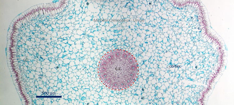
Micrograph of a cross section though an aerial root. The section has been made with a vibratome and stained with safranin – alcian blue. Cellulosic walls are stained blue and lignified/suberized walls are stained pink. As in other roots the section is divided into a small central cylinder (c.c.) and a large cortex.
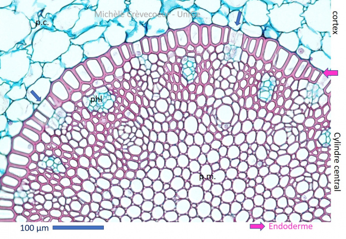
Detailed view of central cylinder with endodermis the innermost layer of cortex characterized by thick walls impregnated with suberin on all their sides. A few passage cells with normal cellulosic unthickened walls are seen (blue arrow) The central cylinder contains xylem and phloem (phl; in blue) distributed in alternance. Phloem appears as small patches of blue stained thin cellulosic walls. Parenchyma cells of the pith are lignified (pm) and those of the cortex (p.c.) are cellulosic.
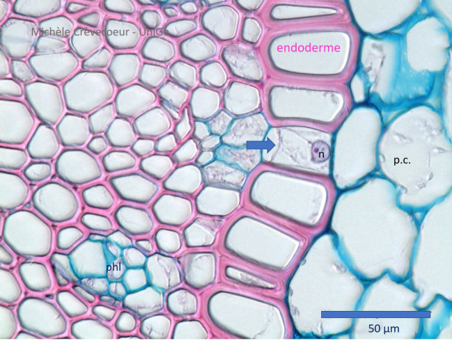
Endodermis at high magnification with living passage cells (blue arrow) in which nucleus is seen (n). All other cells show thick lignified walls (pink stained) on all their faces.
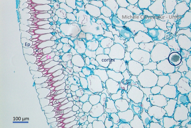
Part of the section illustrating modification of epidermis (ep) with velamen (V) and exodermis (ex). In the cortex cells containing druses are observec (blue circle).
See also micrographs of another section through root of Phalaenopsis stained with astra bleu and basic fuchsin (see protocols).