Microphotographs of longitudinal sections in root of Raphia hookeri palm

Origin of roots: seeds from Côte d’Ivoire germinated and cultured in a multiplication greenhouse in Conservatory and Botanical Garden of the city of Geneva.

The sections have been made by Susanne Mogue Kamga a Master Student in the laboratory of Dr Fred Stauffer – palm Biologist responsible of the Micro Morphology Lab. The sections have been made through roots of the species « Raphia hookeri G. Mann & H. Wendl « . The samples have been embedded in Technovit (see page protocols), sectioned and stained with Toluidine blue.

Micrographs : Michèle Crèvecoeur
Microscope : Leica DM750 equiped with a  caméra MC170HD –  Micromorphology Lab –  Dr Fred Stauffer


Micrographs of a longitudinal section through a root tip and on the left parts of the section showing, the root cap and the meristem (lower part) and cells containing raphides (upper part).

Below high magnification view of cells with  raphides in the upper part of the root on the left.

High magnification view of the section in the differenciation zone with initiation of a lateral root (blue arrow) in front of xylem (x).


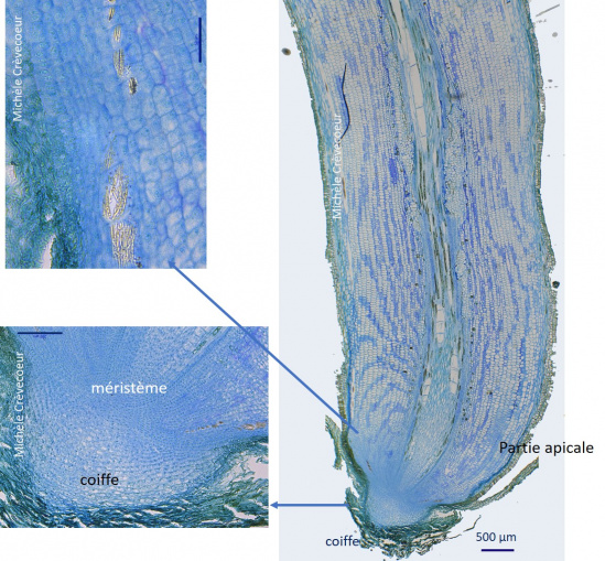
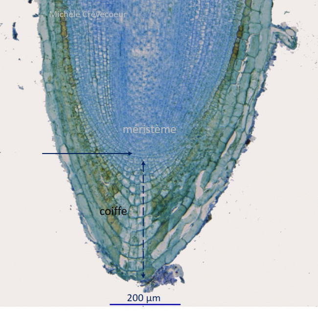
Micrograph of another longitudinal section through a root of raphia palm. The section is more axial and the junction between root cap and meristem is clearly seen (blue arrow). Root cap : dotted blue line.

Site Palmier raphia racine 2.jpg