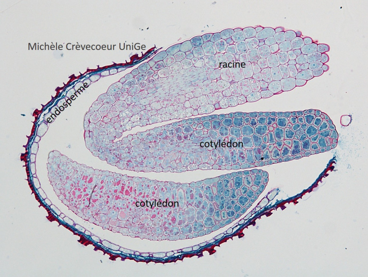
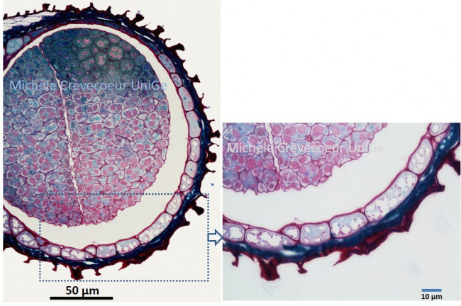
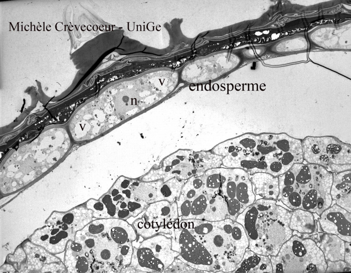
Microphotographs of endosperm in Arabidopsis th. seeds
1/ Light microphotograph of a paraffin section (10 μm)
2/ Light microphotograph of an Epon section (1 micron) stained with methylen blue – basic fuchsin.
3/ Transmission Electron Micrograph of a part of endosperm. The section has been classically stained with lead citrate and uranyle acetate (see protocols).
Higher magnification of the micrograph above.
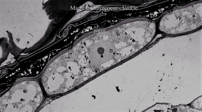
n: nucleus V : vacuoles