DAPI (4′,6-diamidino-2-phenylindole) is a fluorochrome that strongly binds with the minor groove of DNA with a preference for the adenine – thymine base pairs. When bound to double – stranded DNA, DAPI has an absorption maximum at a wavelength of 358 nm (UV) and its emission maximum at 461 nm (blue). It can also bind to RNA probably at the level of adenine – uracil clusters. The DAPI/ RNA complex has an emission maximum at 500 nm a longer wavelength than for the complex DAPI/DNA and with a lower fluorescence.
Protocol for paraffin section (8 μm thick)
Deparaffinize and hydrate to water.
Use a pen to draw a hydrophobic circle around sections
Stain with DAPI at appropriate concentration varying from 0.1 mg/ml to 1 mg/ml.
Add a drop of DAPI on the sections.
Wash 3 x 5 min in distilled water
Mount in appropriating medium such as Vectashied
Observe with a fluorescent microscope using appropriate filters. For Inverted Fluorescent Microscope Leica DM IRE 2 the cube filters used contains an excitation filter in UV (340 – 380 nm); a 400 nm dichroic mirror and an emission filter or barrier filter LP 425 nm.
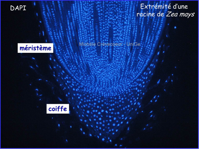
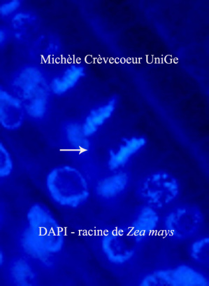
Below : Parts of paraffin sections through a primary Zea mays root tip
Mitosis (white arrow) are clearly seen.
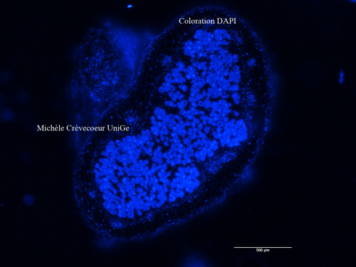
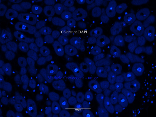
Below : Parts of paraffin sections through root nodules induced by the bacterium strain NGR234