Welcome to Michèle Crèvecoeur’s website dedicated to « plant microscopy »
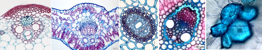
 Passionate about microscopy applied to plant study, I present on this website micrographs made with different types of microscopes as well as protocols for preparing samples to be examined under a microscope in my areas of expertise. The micrographs presented on this site are the results of my experience in these fields since 1975 and specifically the plant world. You will find them in the menu option: Research, Microscopy, Anatomy, Tissues and Protocols. Some micrographs can be found in two different places (anatomy and tissues or anatomy and research ….) and some of them can be found in publications available or not online.
On this site you will also find information about my teaching activities: a list of courses for which I was responsible or co-responsible at the University of Geneva until 2013 as a lecturer, as well as a pdf document which lists the courses, I taught from 1975 to 2010, first in Belgium (University of Liège) and then in Switzerland (Universities of Geneva and Lausanne).
A description of my research activities as a biologist specializing in microscopy at the Department of Botany and Plant Biology of the University of Geneva and an overview of my skills and list of my scientific publications are also presented on this site.
From my 3rd year of scientific training at the University of Liège in Belgium, I was drawn to microscopy and cytology. My master’s thesis and doctorate both defended at this university at the Botanical Institute allowed me to achieve this passion. From 1988 I continued in this path at the University of Geneva. Then in 2003, following a change in my professional status, I set up the « Plant Histology and Cytology Unit » in the Department of Plant Botany and Biology of this University. The unit offered researchers from different research groups of this department responses to their request by proposing very diverse methods of preparing plant samples, making sections and observations with different types of microscopes, photonics, fluorescence, confocal and electronic (see Skills and Microscopy). It also aimed to develop new techniques and approaches according to the researchers needs, to participate in the choice of equipment specific to histology and microscopy within this department and to maintain a collection of anatomical sections in organs, vegetative and floral, of very diverse angiosperms. From 2003, I was helped by Mrs Anne Utz laboratory assistant (25% then 50%), by the many technician apprentices in training and by research and teaching assistants in my laboratory. Students from outside the University of Geneva also came to carry out training courses in my laboratory, among others Mrs Nathalie Jolien within the framework of « Science calls for young people »; Mr François-Xavier Michoud – from the National School of Agronomic Training in Toulouse; Mrs Claire PARENT and Mrs Claire Depardieu – Doctoral students from the University of Franche Comté (Besançon – France).
For more information, please refer to the document: «Research framework from 2000 to 2013″.
Terms of use of this site and its content.
The content of this site is protected by international copyright laws and conventions.
Photo credit : Michèle Crèvecoeur – unige – CH
Micrographs presented on this site can be made available on request.
Site reference : Michèle Crèvecoeur – Plant microscopy.
Date of last update & new pages are indicated below.